
All lanes : Anti-Eg5 antibody (ab37814) at 1/250 dilutionLane 1 : HeLa (Human epithelial carcinoma cell line) Whole Cell LysateLane 2 : Jurkat (Human T cell lymphoblast-like cell line) Whole Cell Lysate (ab7899)Lane 3 : A431 (Human epithelial carcinoma cell line) Whole Cell Lysate (ab7909)Lane 4 : HEK293 (Human embryonic kidney cell line) Whole Cell Lysate (ab7902)Lysates/proteins at 10 µg per lane.SecondaryIRDye 680 Conjugated Goat Anti-Rabbit IgG (H+L) at 1/10000 dilutionPerformed under reducing conditions.
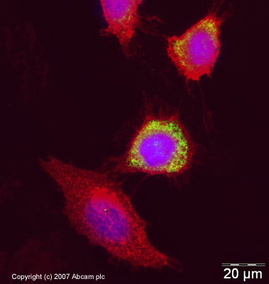
ICC/IF image of ab37814 stained human HeLa cells. The cells were methanol fixed (5 min) and incubated with the antibody (ab37814, 1µg/ml) for 1h at room temperature. The secondary antibody (green) was Alexa Fluor® 488 goat anti-rabbit IgG (H+L) used at a 1/1000 dilution for 1h. Image-iTTM FX Signal Enhancer was used to quench autofluorescence. 5% BSA (in TBS-T) was used for all other blocking steps. DAPI was used to stain the cell nuclei (blue). Alexa Fluor® 594 WGA was used to label plasma membranes (red).
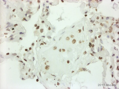
ab37814 (1/250) staining Eg5 in paraffin-embedded Human lung tissue. Tissue underwent fixation in formaldehyde, peroxidase blocking, protein blocking and heat mediated antigen retrieval. The secondary antibody was goat anti rabbit/mouse conjugated to HRP. For further experimental details please refer to abreview.See Abreview