Anti-Eg5 antibody [mAbcam 51976]
| Name | Anti-Eg5 antibody [mAbcam 51976] |
|---|---|
| Supplier | Abcam |
| Catalog | ab51976 |
| Prices | $385.00 |
| Sizes | 100 µg |
| Host | Mouse |
| Clonality | Monoclonal |
| Isotype | IgG1 |
| Clone | mAbcam 51976 |
| Applications | ICC/IF ICC/IF WB IP FC |
| Species Reactivities | Human |
| Antigen | Synthetic peptide conjugated to KLH derived from within residues 950 - 1050 of Human Eg5 |
| Description | Mouse Monoclonal |
| Gene | KIF11 |
| Conjugate | Unconjugated |
| Supplier Page | Shop |
Product images
![All lanes : Anti-Eg5 antibody [mAbcam 51976] (ab51976) at 5 µg/mlLane 1 : HEK293 (Human embryonic kidney cell line) Whole Cell Lysate Lane 2 : A431 (Human epithelial carcinoma cell line) Whole Cell LysateLane 3 : Jurkat (Human T cell lymphoblast-like cell line) Whole Cell Lysate Lysates/proteins at 10 µg per lane.SecondaryRabbit polyclonal to Mouse IgG - H&L (HRP) at 1/2000 dilutionPerformed under reducing conditions.](http://www.bioprodhub.com/system/product_images/ab_products/2/sub_2/12062_ab51976_1_2.jpg) All lanes : Anti-Eg5 antibody [mAbcam 51976] (ab51976) at 5 µg/mlLane 1 : HEK293 (Human embryonic kidney cell line) Whole Cell Lysate Lane 2 : A431 (Human epithelial carcinoma cell line) Whole Cell LysateLane 3 : Jurkat (Human T cell lymphoblast-like cell line) Whole Cell Lysate Lysates/proteins at 10 µg per lane.SecondaryRabbit polyclonal to Mouse IgG - H&L (HRP) at 1/2000 dilutionPerformed under reducing conditions.
All lanes : Anti-Eg5 antibody [mAbcam 51976] (ab51976) at 5 µg/mlLane 1 : HEK293 (Human embryonic kidney cell line) Whole Cell Lysate Lane 2 : A431 (Human epithelial carcinoma cell line) Whole Cell LysateLane 3 : Jurkat (Human T cell lymphoblast-like cell line) Whole Cell Lysate Lysates/proteins at 10 µg per lane.SecondaryRabbit polyclonal to Mouse IgG - H&L (HRP) at 1/2000 dilutionPerformed under reducing conditions.
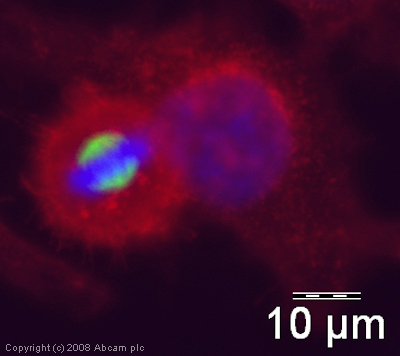 ICC/IF image of ab51976 stained human HeLa cells. The cells were PFA fixed (10 min), permabilised in TBS-T (20 min) and incubated with the antibody (ab51976, 1µg/ml) for 1h at room temperature. 1%BSA / 10% normal goat serum / 0.3M glycine was used to quench autofluorescence and block non-specific protein-protein interactions. The secondary antibody (green) was Alexa Fluor® 488 goat anti-mouse IgG (H+L) used at a 1/1000 dilution for 1h. Alexa Fluor® 594 WGA was used to label plasma membranes (red). DAPI was used to stain the cell nuclei (blue).
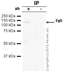 Eg5 was immunoprecipitated using 0.5mg Jurkat whole cell extract, 5µg of Mouse monoclonal to Eg5 and 50µl of protein G magnetic beads (+). No antibody was added to the control (-). The antibody was incubated under agitation with Protein G beads for 10min, Jurkat whole cell extract lysate diluted in RIPA buffer was added to each sample and incubated for a further 10min under agitation.Proteins were eluted by addition of 40µl SDS loading buffer and incubated for 10min at 70oC; 10µl of each sample was separated on a SDS PAGE gel, transferred to a nitrocellulose membrane, blocked with 5% BSA and probed with ab51976.Secondary: Goat polyclonal to mouse IgG light chain specific (HRP) at 1/5000 dilution.Band: 120kDa: Eg5
Product References
APC/C is an essential regulator of centrosome clustering. - APC/C is an essential regulator of centrosome clustering.
Drosopoulos K, Tang C, Chao WC, Linardopoulos S. Nat Commun. 2014 Apr 22;5:3686.
Studies of haspin-depleted cells reveal that spindle-pole integrity in mitosis - Studies of haspin-depleted cells reveal that spindle-pole integrity in mitosis
Dai J, Kateneva AV, Higgins JM. J Cell Sci. 2009 Nov 15;122(Pt 22):4168-76.