![All lanes : Anti-FUBP1 antibody [EPR12326] (ab192867) at 1/1000 dilutionLane 1 : HeLa cell lysateLane 2 : Jurkat cell lysateLane 3 : HepG2 cell lysateLysates/proteins at 10 µg per lane.SecondaryGoat Anti-Rabbit IgG, (H+L), Peroxidase conjugated at 1/1000 dilution](http://www.bioprodhub.com/system/product_images/ab_products/2/sub_2/21220_ab192867-230691-ab1928671.jpg)
All lanes : Anti-FUBP1 antibody [EPR12326] (ab192867) at 1/1000 dilutionLane 1 : HeLa cell lysateLane 2 : Jurkat cell lysateLane 3 : HepG2 cell lysateLysates/proteins at 10 µg per lane.SecondaryGoat Anti-Rabbit IgG, (H+L), Peroxidase conjugated at 1/1000 dilution
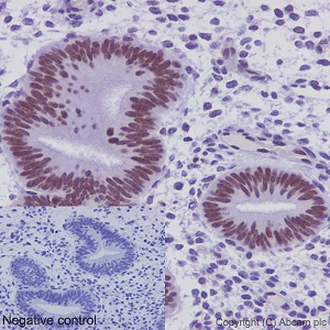
Immunohistochemical analysis of paraffin embedded Human endometrium tissue labeling FUBP1 using ab192867 at a 1/2000 dilution. A prediluted HRP Polymer for Rabbit IgG was used as the secondary antibody. Hematoxylin counterstain.
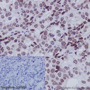
Immunohistochemical analysis of paraffin embedded Human ovarian adenocarcinoma tissue labeling FUBP1 using ab192867 at a 1/2000 dilution. A prediluted HRP Polymer for Rabbit IgG was used as the secondary antibody. Hematoxylin counterstain.
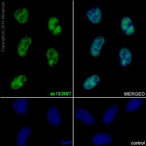
Immunofluorescent analysis of 4% paraformaldehyde fixed HeLa cells labeling FUBP1 using ab192867 at a 1/1000 dilution. A Goat anti rabbit IgG (Alexa Fluor®488) (ab150077) was used as the secondary antibody, at a 1/400 dilution. Counterstain DAPI. Cells were permeabilized using 0.1% Triton X-100.
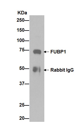
Western blot analysis of pellet from HeLa cell lysate immunoprecipitated using ab192867 at a 1/100 dilution. Secondary antibody was Goat Anti-Rabbit IgG, (H+L), Peroxidase conjugated, at a 1/1000 dilution.