![Standard Curve for G-CSF (Analyte: ab51298) dilution range 1pg/ml to 1ug/ml using Capture Antibody Mouse monoclonal [5D7] to G-CSF (ab9818) at 1ug/ml and Detector Antibody Rabbit polyclonal to G-CSF (ab9691) at 0.5ug/ml](http://www.bioprodhub.com/system/product_images/ab_products/2/sub_2/21607_G-CSF-Primary-antibodies-ab9691-5.jpg)
Standard Curve for G-CSF (Analyte: ab51298) dilution range 1pg/ml to 1ug/ml using Capture Antibody Mouse monoclonal [5D7] to G-CSF (ab9818) at 1ug/ml and Detector Antibody Rabbit polyclonal to G-CSF (ab9691) at 0.5ug/ml
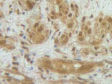
ab9691 staining G-CSF in human angiosarcoma section by Immunohistochemistry (Formalin/PFA fixed paraffin-embedded sections). Tissue underwent heat mediated antigen retrieval in sodium citrate buffer (pH 6.0). The primary antibody was used at 0.25 ug/ml and incubated with sample at 4°C overnight. A HRP-labeled polymer detection system was used with a DAB chromogen.
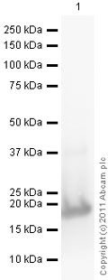
developed using the ECL techniquePerformed under reducing conditions.
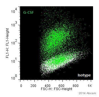
ab9691 staining G-CSF in Human HL-1 cells by Flow Cytometry. Cells were fixed with paraformaldehyde and permeabilized. The sample was incubated with the primary antibody (1/300) for 1 hour at 20°C. An Alexa Fluor® 488-conjugated goat anti-rabbit IgG (1/1000) was used as the secondary antibody.Gating Strategy: Isotype population (shown in white).See Abreview