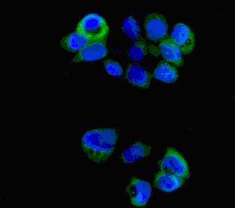
Immunofluorescent analysis of HT-1376 cells (paraformaldehyde-fixed, 4%) labeling G-CSF with ab181053 at 1/100 dilution followed by Goat anti rabbit IgG (Alexa Fluor® 488) secondary at 1/200 dilution and counter-stained with DAPI (blue).
![All lanes : Anti-G-CSF antibody [EPR3203(N)(B)] (ab181053) at 1/2000 dilutionLane 1 : K562 cell lysateLane 2 : KM3 cell lysateLane 3 : NCI-H460 cell lysateLane 4 : HT-1376 cell lysateLysates/proteins at 20 µg per lane.SecondaryGoat Anti-Rabbit IgG, (H+L) HRP at 1/1000 dilution](http://www.bioprodhub.com/system/product_images/ab_products/2/sub_2/21618_ab181053-213069-ab181053.jpg)
All lanes : Anti-G-CSF antibody [EPR3203(N)(B)] (ab181053) at 1/2000 dilutionLane 1 : K562 cell lysateLane 2 : KM3 cell lysateLane 3 : NCI-H460 cell lysateLane 4 : HT-1376 cell lysateLysates/proteins at 20 µg per lane.SecondaryGoat Anti-Rabbit IgG, (H+L) HRP at 1/1000 dilution
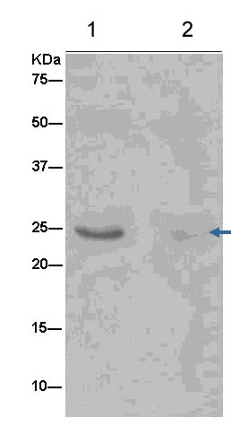
Western blot analysis of immunoprecipitation pellet from K562 cell lysate (lane 1) or a Negative control (lane 2) immunoprecipitated using ab181053 at 1/20 dilution.Secondary: Anti-Rabbit IgG (HRP), specific to the non-reduced form of IgG at 1/1500 dilution.