![All lanes : Anti-GRB2 antibody [Y301] (ab32111) at 1/2000 dilutionLane 1 : NIH 3T3 cell lysateLane 2 : PC12 cell lysateLane 3 : HeLa cell lysate](http://www.bioprodhub.com/system/product_images/ab_products/2/sub_2/27492_WB%2520GRB2.jpg)
All lanes : Anti-GRB2 antibody [Y301] (ab32111) at 1/2000 dilutionLane 1 : NIH 3T3 cell lysateLane 2 : PC12 cell lysateLane 3 : HeLa cell lysate
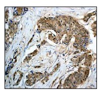
Immunohistochemical analysis of GRB2 expression in paraffin embedded human breast carcinoma tissue section, using 1/100 ab32111.
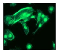
Immunofluorescent analysis of GRB2 expression in PC12 cells, using 1/100 ab32111.
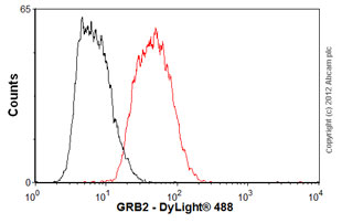
Overlay histogram showing SH-SY5Y cells stained with ab32111 (red line). The cells were fixed with 4% paraformaldehyde (10 min) and then permeabilized with 0.1% PBS-Tween for 20 min. The cells were then incubated in 1x PBS / 10% normal goat serum / 0.3M glycine to block non-specific protein-protein interactions followed by the antibody (ab32111, 1/100 dilution) for 30 min at 22ºC. The secondary antibody used was DyLight® 488 goat anti-rabbit IgG (H+L) (ab96899) at 1/500 dilution for 30 min at 22ºC. Isotype control antibody (black line) was rabbit IgG (monoclonal) (1µg/1x106 cells) used under the same conditions. Acquisition of >5,000 events was performed.