Anti-GRP78 antibody (ab188878) at 1/1000 dilution + Mouse liver lysate
Confocal immunofluorescent analysis of NCI H460 cells labeling GRP78 with ab188878 at 1/10 dilution followed by Alexa Fluor 488-conjugated goat anti-rabbit lgG (green). DAPI was used to stain the cell nuclear (blue).
Anti-GRP78 antibody (ab188878) at 1/1000 dilution + HL60 cell lysate at 35 µg
Flow cytometric analysis of HeLa cells labeling GRP78 with ab188878 at 1/10 dilution (right histogram) compared with a negative control cell (left) followed by FITC-conjugated goat-anti-rabbit secondary antibody.
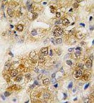
Immunohistochemical analysis of formalin-fixed, paraffin-embedded Human prostate carcinoma tissue labeling GRP78 with ab188878 at 10 µg/ml followed by peroxidase-conjugated secondary antibody and DAB staining.
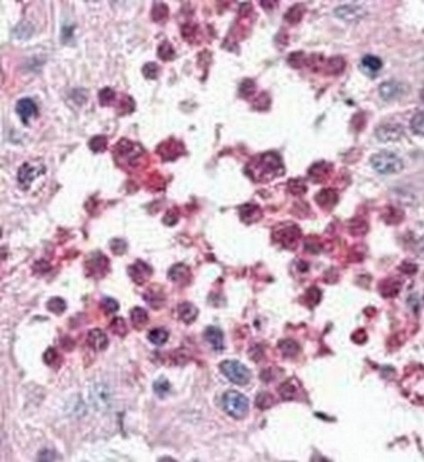
Immunohistochemical analysis of formalin-fixed, paraffin-embedded Human testis tissue labeling GRP78 with ab188878 at 10 µg/ml followed by peroxidase-conjugated secondary antibody and AEC staining.
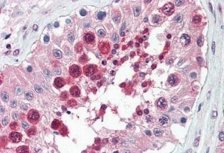
Immunohistochemical analysis of formalin-fixed, paraffin-embedded Human testis tissue labeling GRP78 with ab188878 at 10 µg/ml followed by biotinylated secondary antibody, alkaline phosphatase-streptavidin and chromogen.
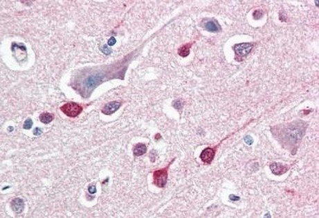
Immunohistochemical analysis of formalin-fixed, paraffin-embedded Human brain cortex tissue labeling GRP78 with ab188878 at 10 µg/ml followed by biotinylated secondary antibody, alkaline phosphatase-streptavidin and chromogen.