Anti-HDAC1 antibody
| Name | Anti-HDAC1 antibody |
|---|---|
| Supplier | Abcam |
| Catalog | ab33278 |
| Prices | $398.00 |
| Sizes | 100 µg |
| Host | Rabbit |
| Clonality | Polyclonal |
| Isotype | IgG |
| Applications | IHC-F WB ICC/IF ICC/IF |
| Species Reactivities | Rat, Human, Xenopus, Zebrafish, Mouse, Chicken, Bovine |
| Antigen | Synthetic peptide conjugated to KLH derived from within residues 50 - 150 of Zebrafish HDAC1 |
| Description | Rabbit Polyclonal |
| Gene | HDAC1 |
| Conjugate | Unconjugated |
| Supplier Page | Shop |
Product images
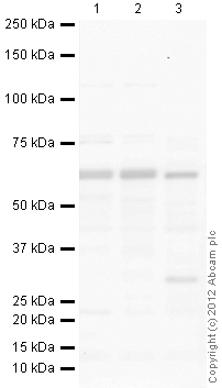 All lanes : Anti-HDAC1 antibody (ab33278) at 1 µg/mlLane 1 : HeLa (Human epithelial carcinoma cell line) Whole Cell LysateLane 2 : HEK293 (Human embryonic kidney cell line) Whole Cell LysateLane 3 : Zebrafish Embryo ExtractLysates/proteins at 10 µg per lane.SecondaryGoat Anti-Rabbit IgG H&L (HRP) preadsorbed (ab97080) at 1/5000 dilutiondeveloped using the ECL techniquePerformed under reducing conditions.
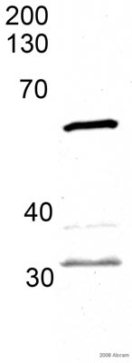 Anti-HDAC1 antibody (ab33278) at 0.06 µg/ml + whole embryo zebrafish tissue at 45 µgSecondarywhole embryo zebrafish tissue lysate at 1/5000 dilution
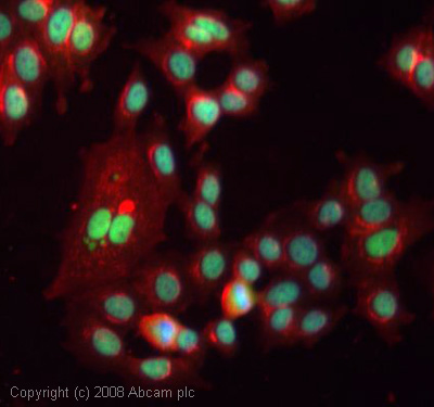 ICC/IF image of ab33278 stained human MCF7 cells. The cells were methanol fixed (5 min), permabilised in 0.1% PBS-Tween (20 min) and incubated with the antibody (ab33278, 1µg/ml) for 1h at room temperature. 1%BSA / 10% normal goat serum / 0.3M glycine was used to block non-specific protein-protein interactions. The secondary antibody (green) was Alexa Fluor® 488 goat anti-rabbit IgG (H+L) used at a 1/1000 dilution for 1h. Alexa Fluor® 594 WGA was used to label plasma membranes (red). DAPI was used to stain the cell nuclei (blue). This antibody also gave a positive IF result in HeLa, HEK 293 and HepG2 cells.
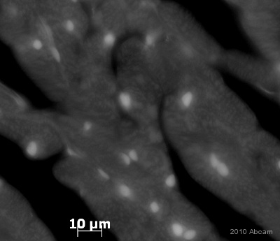 Immunohistochemistical detection of HDAC1 using antibody ab33278 on PFA perfusion fixed frozen rat heart sections. Primary antibody Dilution 1/300, Incubated for 18 hours @ 20°C in PBS + 0.3 % Triton X100. Secondary antibody: Goat anti-rabbit Alexa Fluor 488 (1/1000). The antibody produced some staining in the cytoplasm and what seems to be the nucleus of muscle cells in rat heart tissue. The tissues were fixed (animals perfused fixed) with 4% PFA and later postfixed overnight in the same fixative. They were cryoprotected in 30% sucrose and cut using a cryostat.See Abreview
Product References
Inhibition of endothelial ERK signalling by Smad1/5 is essential for - Inhibition of endothelial ERK signalling by Smad1/5 is essential for
Zhang C, Lv J, He Q, Wang S, Gao Y, Meng A, Yang X, Liu F. Nat Commun. 2014 Mar 11;5:3431.
Histone deacetylase expression patterns in developing murine optic nerve. - Histone deacetylase expression patterns in developing murine optic nerve.
Tiwari S, Dharmarajan S, Shivanna M, Otteson DC, Belecky-Adams TL. BMC Dev Biol. 2014 Jul 9;14:30.
Distinct functional and temporal requirements for zebrafish Hdac1 during neural - Distinct functional and temporal requirements for zebrafish Hdac1 during neural
Ignatius MS, Unal Eroglu A, Malireddy S, Gallagher G, Nambiar RM, Henion PD. PLoS One. 2013 May 7;8(5):e63218.