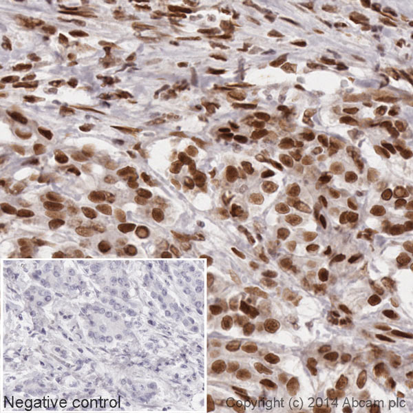
IHC image of HDAC1 staining in a section of formalin-fixed paraffin-embedded normal human pancreas*. The section was pre-treated using pressure cooker heat mediated antigen retrieval with sodium citrate buffer (pH6) for 30 mins. The section was incubated with ab193096 at a working dilution of 1 in 500 overnight at +4°C. The section was counterstained with haematoxylin and mounted with DPX.The inset negative control image is taken from an identical assay without primary antibody.For other IHC staining systems (automated and non-automated) customers should optimize variable parameters such as antigen retrieval conditions, primary antibody concentration and antibody incubation times.*Tissue obtained from the Human Research Tissue Bank, supported by the NIHR Cambridge Biomedical Research Centre
![All lanes : Anti-HDAC1 antibody [EPR460(2)] (HRP) (ab193096) at 1/5000 dilutionLane 1 : Jurkat (Human T cell lymphoblast-like cell line) Whole Cell LysateLane 2 : HeLa (Human epithelial carcinoma cell line) Whole Cell Lysate (ab150035)Lane 3 : K562 (Human erythromyeloblastoid leukemia cell line) Whole Cell LysateLane 4 : MCF7 (Human breast adenocarcinoma cell line) Whole Cell LysateLysates/proteins at 10 µg per lane.developed using the ECL techniquePerformed under reducing conditions.](http://www.bioprodhub.com/system/product_images/ab_products/2/sub_2/29269_ab193096-232638-WBAP20888701.jpg)
All lanes : Anti-HDAC1 antibody [EPR460(2)] (HRP) (ab193096) at 1/5000 dilutionLane 1 : Jurkat (Human T cell lymphoblast-like cell line) Whole Cell LysateLane 2 : HeLa (Human epithelial carcinoma cell line) Whole Cell Lysate (ab150035)Lane 3 : K562 (Human erythromyeloblastoid leukemia cell line) Whole Cell LysateLane 4 : MCF7 (Human breast adenocarcinoma cell line) Whole Cell LysateLysates/proteins at 10 µg per lane.developed using the ECL techniquePerformed under reducing conditions.