Anti-HIBADH antibody [EPR12519(B)]
| Name | Anti-HIBADH antibody [EPR12519(B)] |
|---|---|
| Supplier | Abcam |
| Catalog | ab175203 |
| Prices | $357.00 |
| Sizes | 100 µl |
| Host | Rabbit |
| Clonality | Monoclonal |
| Isotype | IgG |
| Clone | EPR12519(B) |
| Applications | IHC-P WB ICC/IF ICC/IF IP FC |
| Species Reactivities | Human, Bovine, Orangutan |
| Antigen | Synthetic peptide (the amino acid sequence is considered to be commercially sensitive) within Human HIBADH aa 50-150 (Cysteine residue) |
| Description | Rabbit Monoclonal |
| Gene | HIBADH |
| Conjugate | Unconjugated |
| Supplier Page | Shop |
Product images
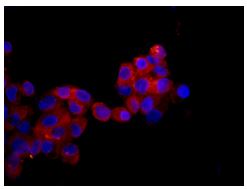 Immunofluorescent staining of HepG2 cells labeling HIBADH with ab175203 at 1/100 dilution (red). DAPI nuclear staining (blue).
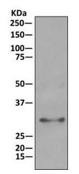
![All lanes : Anti-HIBADH antibody [EPR12519(B)] (ab175203) at 1/1000 dilutionLane 1 : Human fetal liver tissue lysateLane 2 : Human fetal heart tissue lysateLane 3 : HepG2 cell lysateLane 4 : Human fetal kidney tissue lysateLysates/proteins at 10 µg per lane.](http://www.bioprodhub.com/system/product_images/ab_products/2/sub_3/472_ab175203-201694-ab1752031.JPG) All lanes : Anti-HIBADH antibody [EPR12519(B)] (ab175203) at 1/1000 dilutionLane 1 : Human fetal liver tissue lysateLane 2 : Human fetal heart tissue lysateLane 3 : HepG2 cell lysateLane 4 : Human fetal kidney tissue lysateLysates/proteins at 10 µg per lane.
All lanes : Anti-HIBADH antibody [EPR12519(B)] (ab175203) at 1/1000 dilutionLane 1 : Human fetal liver tissue lysateLane 2 : Human fetal heart tissue lysateLane 3 : HepG2 cell lysateLane 4 : Human fetal kidney tissue lysateLysates/proteins at 10 µg per lane.
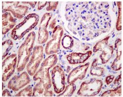 Immunohistochemical analysis of paraffin embedded Human kidney tissue labeling HIBADH with ab175203 at 1/50 dilution.
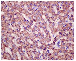 Immunohistochemical analysis of paraffin embedded Human liver tissue labeling HIBADH with ab175203 at 1/50 dilution.
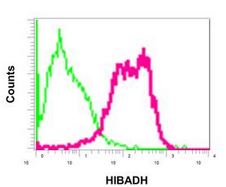 Flow cytometric analysis of permeabilized HepG2 cells labeling HIBADH with ab175203 at 1/10 dilution (red) compared to a Rabbit IgG (green).
 Western blot analysis on immunoprecipitation pellet from HepG2 cell lysate labeling HIBADH with ab175203 at 1/10 dilution.