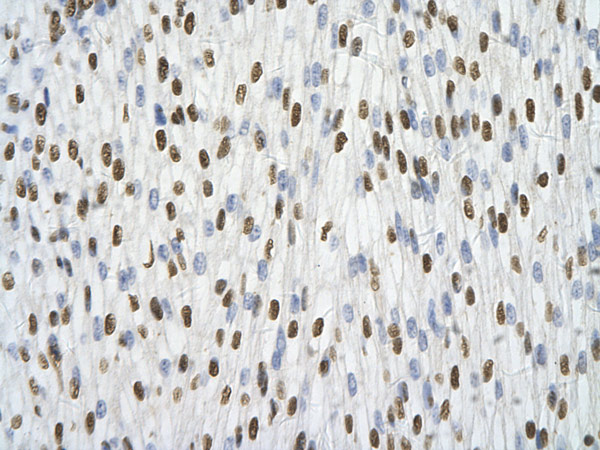
Immunohistochemistry (Formalin/PFA-fixed paraffin-embedded sections) analysis of human heart tissue labelling hnRNP A1 with ab50492 at 4.0-8.0µg/ml. Positive staining shown in cardiac cell. Magnification: 400X.
Ab50492, at a concentration of 4 - 8 µg/ml, staining hnRNP A1 in paraffin embedded human liver cells by Immunohistochemistry. Hepatocytes cells are positively stained (arrows). Magnification 400x.Ab50492, at a concentration of 4 - 8 µg/ml, staining hnRNP A1 in paraffin embedded human liver cells by Immunohistochemistry. Hepatocytes cells are positively stained (arrows). Magnification 400x.
Ab50492, at a concentration of 4 - 8 µg/ml, staining hnRNP A1 in paraffin embedded human heart tissue by Immunohistochemistry. Myocardial cells are positively stained (arrows). Magnification 400x.Ab50492, at a concentration of 4 - 8 µg/ml, staining hnRNP A1 in paraffin embedded human heart tissue by Immunohistochemistry. Myocardial cells are positively stained (arrows). Magnification 400x.
Lane 1 : MarkerLane 2 : Anti-hnRNP A1 antibody (ab50492) at 1.25 µg/mlLane 1 : As aboveLane 2 : Jurkat cell lysateSecondaryHRP conjugated anti-Rabbit IgG at 1/50000 dilution
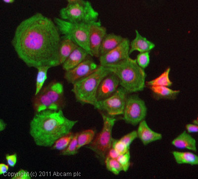
ICC/IF image of ab50492 stained MCF7 cells. The cells were 4% formaldehyde fixed (10 min) and then incubated in 1%BSA / 10% normal goat serum / 0.3M glycine in 0.1% PBS-Tween for 1h to permeabilise the cells and block non-specific protein-protein interactions. The cells were then incubated with the antibody (ab50492, 5µg/ml) overnight at +4°C. The secondary antibody (green) was ab96899, DyLight® 488 goat anti-rabbit IgG (H+L) used at a 1/250 dilution for 1h. Alexa Fluor® 594 WGA was used to label plasma membranes (red) at a 1/200 dilution for 1h. DAPI was used to stain the cell nuclei (blue) at a concentration of 1.43µM.