Anti-Hsp60 antibody [LK-1]
| Name | Anti-Hsp60 antibody [LK-1] |
|---|---|
| Supplier | Abcam |
| Catalog | ab59457 |
| Prices | $403.00 |
| Sizes | 100 µg |
| Host | Mouse |
| Clonality | Monoclonal |
| Isotype | IgG1 |
| Clone | LK-1 |
| Applications | WB IP ELISA FC IHC-F IHC-P |
| Species Reactivities | Mouse, Rat, Sheep, Rabbit, Chicken, Guinea Pig, Hamster, Bovine, Dog, Human, Pig, Xenopus, Drosophila, Monkey |
| Antigen | Human Hsp60 produced through recombinant DNA methods in E |
| Description | Mouse Monoclonal |
| Gene | HSPD1 |
| Conjugate | Unconjugated |
| Supplier Page | Shop |
Product images
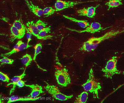 ICC/IF image of ab59457 stained HepG2 cells. The cells were 100% methanol fixed (5 min) and then incubated in 1%BSA / 10% normal goat serum / 0.3M glycine in 0.1% PBS-Tween for 1h to permeabilise the cells and block non-specific protein-protein interactions. The cells were then incubated with the antibody (ab59457, 1µg/ml) overnight at +4°C. The secondary antibody (green) was Alexa Fluor® 488 goat anti-mouse IgG (H+L) used at a 1/1000 dilution for 1h. Alexa Fluor® 594 WGA was used to label plasma membranes (red) at a 1/200 dilution for 1h. DAPI was used to stain the cell nuclei (blue) at a concentration of 1.43µM.
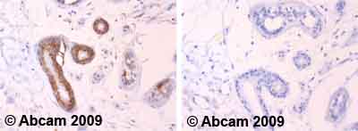 Ab59457 staining human normal skin tissue. Staining is localised to mitochondria.Left panel: with primary antibody at 2 ug/ml. Right panel: isotype control.Sections were stained using an automated system DAKO Autostainer Plus , at room temperature. Sections were rehydrated and antigen retrieved with the Dako 3-in-1 antigen retrieval buffer EDTA pH 9.0 in a DAKO PT Link. Slides were peroxidase blocked in 3% H2O2 in methanol for 10 minutes. They were then blocked with Dako Protein block for 10 minutes (containing casein 0.25% in PBS) then incubated with primary antibody for 20 minutes and detected with Dako Envision Flex amplification kit for 30 minutes. Colorimetric detection was completed with diaminobenzidine for 5 minutes. Slides were counterstained with Haematoxylin and coverslipped under DePeX. Please note that for manual staining we recommend to optimize the primary antibody concentration and incubation time (overnight incubation), and amplification may be required.
![All lanes : Anti-Hsp60 antibody [LK-1] (ab59457) at 0.05 µg/mlLane 1 : Rat Brain tissue lysatesLane 2 : Rat Heart tissue lysatesLane 3 : Rat Kidney tissue lysatesLane 4 : Rat Liver tissue lysatesLane 5 : Rat Lung tissue lysatesLane 6 : Rat Pancreas tissue lysatesLane 7 : Rat skeletal muscle tissue lysateLane 8 : Rat Spleen tissue lysateLane 9 : Rat Testes tissue lysateLane 10 : Rat Thymus tisuue lysateLane 11 : Cell lysates prepared from rat heart H9C2 cellsLane 12 : Cell lysates prepared from mouse NIH3T3 cellsLane 13 : Cell lysates prepared from mouse Pam212 cellsLysates/proteins at 10 µg per lane.SecondaryHRP-conjugated goat polyclonal to mouse IgG1 at 1/10 dilution](http://www.bioprodhub.com/system/product_images/ab_products/2/sub_3/4963_Hsp60-Primary-antibodies-ab59457-3.jpg) All lanes : Anti-Hsp60 antibody [LK-1] (ab59457) at 0.05 µg/mlLane 1 : Rat Brain tissue lysatesLane 2 : Rat Heart tissue lysatesLane 3 : Rat Kidney tissue lysatesLane 4 : Rat Liver tissue lysatesLane 5 : Rat Lung tissue lysatesLane 6 : Rat Pancreas tissue lysatesLane 7 : Rat skeletal muscle tissue lysateLane 8 : Rat Spleen tissue lysateLane 9 : Rat Testes tissue lysateLane 10 : Rat Thymus tisuue lysateLane 11 : Cell lysates prepared from rat heart H9C2 cellsLane 12 : Cell lysates prepared from mouse NIH3T3 cellsLane 13 : Cell lysates prepared from mouse Pam212 cellsLysates/proteins at 10 µg per lane.SecondaryHRP-conjugated goat polyclonal to mouse IgG1 at 1/10 dilution
All lanes : Anti-Hsp60 antibody [LK-1] (ab59457) at 0.05 µg/mlLane 1 : Rat Brain tissue lysatesLane 2 : Rat Heart tissue lysatesLane 3 : Rat Kidney tissue lysatesLane 4 : Rat Liver tissue lysatesLane 5 : Rat Lung tissue lysatesLane 6 : Rat Pancreas tissue lysatesLane 7 : Rat skeletal muscle tissue lysateLane 8 : Rat Spleen tissue lysateLane 9 : Rat Testes tissue lysateLane 10 : Rat Thymus tisuue lysateLane 11 : Cell lysates prepared from rat heart H9C2 cellsLane 12 : Cell lysates prepared from mouse NIH3T3 cellsLane 13 : Cell lysates prepared from mouse Pam212 cellsLysates/proteins at 10 µg per lane.SecondaryHRP-conjugated goat polyclonal to mouse IgG1 at 1/10 dilution
![Overlay histogram showing HeLa cells stained with ab59457 (red line). The cells were fixed with 80% methanol (5 min) and then permeabilized with 0.1% PBS-Tween for 20 min. The cells were then incubated in 1x PBS / 10% normal goat serum / 0.3M glycine to block non-specific protein-protein interactions followed by the antibody (ab59457, 1µg/1x106 cells) for 30 min at 22ºC. The secondary antibody used was DyLight® 488 goat anti-mouse IgG (H+L) (ab96879) at 1/500 dilution for 30 min at 22ºC. Isotype control antibody (black line) was mouse IgG1 [ICIGG1] (ab91353, 2µg/1x106 cells) used under the same conditions. Acquisition of >5,000 events was performed.](http://www.bioprodhub.com/system/product_images/ab_products/2/sub_3/4964_Hsp60-Primary-antibodies-ab59457-8.jpg) Overlay histogram showing HeLa cells stained with ab59457 (red line). The cells were fixed with 80% methanol (5 min) and then permeabilized with 0.1% PBS-Tween for 20 min. The cells were then incubated in 1x PBS / 10% normal goat serum / 0.3M glycine to block non-specific protein-protein interactions followed by the antibody (ab59457, 1µg/1x106 cells) for 30 min at 22ºC. The secondary antibody used was DyLight® 488 goat anti-mouse IgG (H+L) (ab96879) at 1/500 dilution for 30 min at 22ºC. Isotype control antibody (black line) was mouse IgG1 [ICIGG1] (ab91353, 2µg/1x106 cells) used under the same conditions. Acquisition of >5,000 events was performed.
Overlay histogram showing HeLa cells stained with ab59457 (red line). The cells were fixed with 80% methanol (5 min) and then permeabilized with 0.1% PBS-Tween for 20 min. The cells were then incubated in 1x PBS / 10% normal goat serum / 0.3M glycine to block non-specific protein-protein interactions followed by the antibody (ab59457, 1µg/1x106 cells) for 30 min at 22ºC. The secondary antibody used was DyLight® 488 goat anti-mouse IgG (H+L) (ab96879) at 1/500 dilution for 30 min at 22ºC. Isotype control antibody (black line) was mouse IgG1 [ICIGG1] (ab91353, 2µg/1x106 cells) used under the same conditions. Acquisition of >5,000 events was performed.
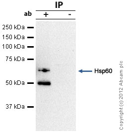 Hsp60 was immunoprecipitated using 0.5mg Rat Brain tissue lysate, 5µg of Mouse monoclonal to Hsp60 and 50µl of protein G magnetic beads (+). No antibody was added to the control (-). The antibody was incubated under agitation with Protein G beads for 10min, Rat Brain tissue lysate lysate diluted in RIPA buffer was added to each sample and incubated for a further 10min under agitation.Proteins were eluted by addition of 40µl SDS loading buffer and incubated for 10min at 70oC; 10µl of each sample was separated on a SDS PAGE gel, transferred to a nitrocellulose membrane, blocked with 5% BSA and probed with ab59457.Secondary: Goat polyclonal to mouse IgG light chain specific (HRP) at 1/20,000 dilution.Band: 60kDa; Hsp60: non specific bands - 50kDa: We are unsure as to the identity of this extra band.
Product References
Uncoupling protein 2 and 4 expression pattern during stem cell differentiation - Uncoupling protein 2 and 4 expression pattern during stem cell differentiation
Rupprecht A, Sittner D, Smorodchenko A, Hilse KE, Goyn J, Moldzio R, Seiler AE, Brauer AU, Pohl EE. PLoS One. 2014 Feb 11;9(2):e88474.
Mitophagy-dependent necroptosis contributes to the pathogenesis of COPD. - Mitophagy-dependent necroptosis contributes to the pathogenesis of COPD.
Mizumura K, Cloonan SM, Nakahira K, Bhashyam AR, Cervo M, Kitada T, Glass K, Owen CA, Mahmood A, Washko GR, Hashimoto S, Ryter SW, Choi AM. J Clin Invest. 2014 Sep;124(9):3987-4003.
Elevated mitochondrial oxidative stress impairs metabolic adaptations to exercise - Elevated mitochondrial oxidative stress impairs metabolic adaptations to exercise
Crane JD, Abadi A, Hettinga BP, Ogborn DI, MacNeil LG, Steinberg GR, Tarnopolsky MA. PLoS One. 2013 Dec 6;8(12):e81879.
The C8ORF38 homologue Sicily is a cytosolic chaperone for a mitochondrial complex - The C8ORF38 homologue Sicily is a cytosolic chaperone for a mitochondrial complex
Zhang K, Li Z, Jaiswal M, Bayat V, Xiong B, Sandoval H, Charng WL, David G, Haueter C, Yamamoto S, Graham BH, Bellen HJ. J Cell Biol. 2013 Mar 18;200(6):807-20.