![All lanes : Anti-MAP1LC3A antibody [166AT1234] (ab168803) at 1/1000 dilutionLane 1 : Hela cell lysates treated with DMSO Lane 2 : Hela cell lysates treated with rapamycin Lane 3 : Hela cell lysates treated with bafilomycin Lysates/proteins at 12.5 µg per lane.](http://www.bioprodhub.com/system/product_images/ab_products/2/sub_3/19181_MAP1LC3A-Primary-antibodies-ab168803-4.jpg)
All lanes : Anti-MAP1LC3A antibody [166AT1234] (ab168803) at 1/1000 dilutionLane 1 : Hela cell lysates treated with DMSO Lane 2 : Hela cell lysates treated with rapamycin Lane 3 : Hela cell lysates treated with bafilomycin Lysates/proteins at 12.5 µg per lane.
![All lanes : Anti-MAP1LC3A antibody [166AT1234] (ab168803) at 8 µg/mlLane 1 : Y79 (soluble fraction of cell extract)Lane 2 : 293 transfected with human MAP1LC3A (whole cell extract)Lysates/proteins at 12.5 µg per lane.](http://www.bioprodhub.com/system/product_images/ab_products/2/sub_3/19182_MAP1LC3A-Primary-antibodies-ab168803-6.jpg)
All lanes : Anti-MAP1LC3A antibody [166AT1234] (ab168803) at 8 µg/mlLane 1 : Y79 (soluble fraction of cell extract)Lane 2 : 293 transfected with human MAP1LC3A (whole cell extract)Lysates/proteins at 12.5 µg per lane.
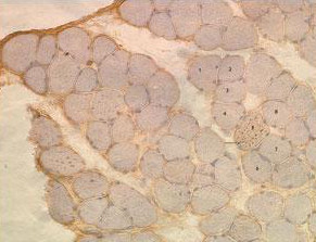
Immunohistochemistry analysis of muscle tissue of a diseased Mouse off Dox after 5 weeks on regular food CD2 labeling MAP1LC3A with ab168803 at 1/50.
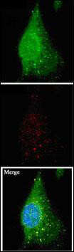
Immunocytochemistry analysis of Mouse cerebellar cell lines stably expressing Human GFP-MAP1LC3A fusion protein labeling MAP1LC3A with ab168803 at 1/10. Conditions: 1:1 methanol:acetone fixation, 0.2% saponin permeabilization.
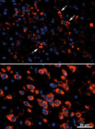
Immunocytochemistry analysis of Cathepsin D WT (bottom) and KO (top) Mouse thalamus (postnatal day 25) labeling MAP1LC3A with ab168803 at 1/10.