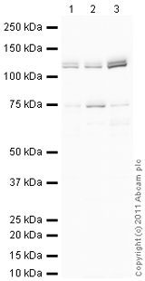
All lanes : Anti-Nicastrin antibody (ab122969) at 1 µg/mlLane 1 : E10 Mouse Embryo Brain Tissue LysateLane 2 : E12 Mouse Embryo Brain and Spinal Cord Tissue LysateLane 3 : E16 Mouse Embryo Brain Tissue LysateLysates/proteins at 10 µg per lane.SecondaryGoat Anti-Rabbit IgG H&L (HRP) preadsorbed (ab97080) at 1/5000 dilutiondeveloped using the ECL techniquePerformed under reducing conditions.
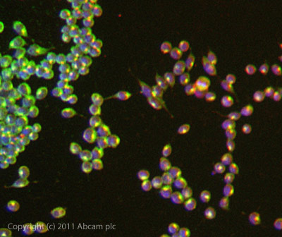
ICC/IF image of ab122969 stained Mouse Embryonic Stem cells. The cells were 100% methanol fixed (5 min) and then incubated in 1%BSA / 10% normal goat serum / 0.3M glycine in 0.1% PBS-Tween for 1h to permeabilise the cells and block non-specific protein-protein interactions. The cells were then incubated with the antibody ab122969 at 5µg/ml overnight at +4°C. The secondary antibody (green) was DyLight® 488 goat anti- rabbit (ab96899) IgG (H+L) used at a 1/250 dilution for 1h. Alexa Fluor® 594 WGA was used to label plasma membranes (red) at a 1/200 dilution for 1h. DAPI was used to stain the cell nuclei (blue) at a concentration of 1.43µM.
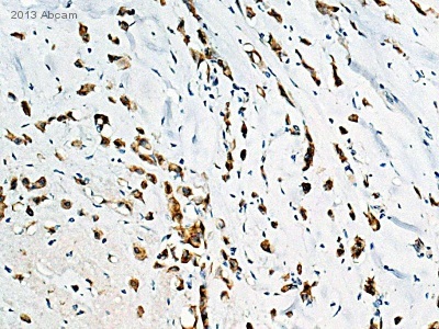
ab122969 staining Nicastrin in Human breast carcinoma tissue sections by Immunohistochemistry (IHC-P - paraformaldehyde-fixed, paraffin-embedded sections). Tissue was fixed with formaldehyde and blocked with 1% BSA for 10 minutes at 21°C; antigen retrieval was by heat mediation in a citrate buffer. Samples were incubated with primary antibody (1/8000 in TBS/BSA/azide) for 16 hours at 21°C. A Biotin-conjugated Goat anti-rabbit IgG polyclonal (1/250) was used as the secondary antibody.See Abreview