![All lanes : Anti-NTH1 antibody [2660C1a] (ab70726) at 0.2 µg/mlLane 1 : Whole cell lysate from human HEK293 cellsLane 2 : Whole cell lysate from mouse NIH3T3 cellsLane 3 : Whole cell lysate from rat F2408 cellsLysates/proteins at 25 µg per lane.](http://www.bioprodhub.com/system/product_images/ab_products/2/sub_4/2447_ab70726-2.jpg)
All lanes : Anti-NTH1 antibody [2660C1a] (ab70726) at 0.2 µg/mlLane 1 : Whole cell lysate from human HEK293 cellsLane 2 : Whole cell lysate from mouse NIH3T3 cellsLane 3 : Whole cell lysate from rat F2408 cellsLysates/proteins at 25 µg per lane.
![Anti-NTH1 antibody [2660C1a] (ab70726) at 0.2 µg/ml + immunised recombinant human NTH1 protein](http://www.bioprodhub.com/system/product_images/ab_products/2/sub_4/2448_ab70726.gif)
Anti-NTH1 antibody [2660C1a] (ab70726) at 0.2 µg/ml + immunised recombinant human NTH1 protein
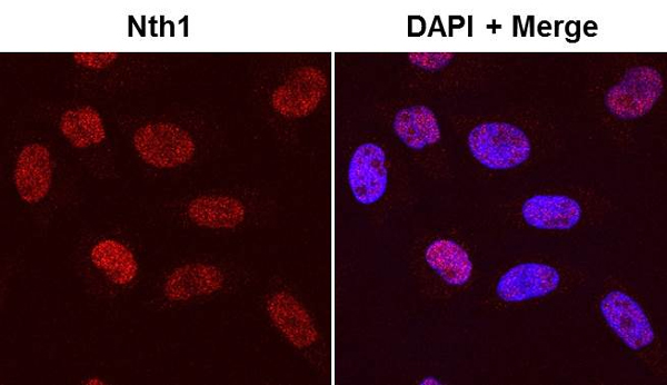
ab70726 staining NTH1 in HeLa cells by ICC/IF (Immunocytochemistry/immunofluorescence). Cells were fixed with paraformaldehyde, permeabilized with 0.2% Triton X-100 and blocked with 2% BSA for 45 minutes at room temperature. Samples were incubated with primary antibody (1/300 in PBS + 2% BSA) for 14 hours at 4°C. An Alexa Fluor® 594-conjugated goat anti-mouse IgG polyclonal (1/500) was used as the secondary antibody.See Abreview