Anti-NUMBL antibody
| Name | Anti-NUMBL antibody |
|---|---|
| Supplier | Abcam |
| Catalog | ab37500 |
| Prices | $370.00 |
| Sizes | 100 µg |
| Host | Rabbit |
| Clonality | Polyclonal |
| Isotype | IgG |
| Applications | WB ICC/IF ICC/IF IHC-F IHC-F |
| Species Reactivities | Mouse, Rat, Bovine, Human |
| Antigen | Synthetic peptide conjugated to KLH derived from within residues 200 - 300 of Human NUMBL |
| Description | Rabbit Polyclonal |
| Gene | NUMBL |
| Conjugate | Unconjugated |
| Supplier Page | Shop |
Product images
 All lanes : Anti-NUMBL antibody (ab37500) at 1 µg/mlLane 1 : Brain (Mouse) Whole Cell Lysate - normal tissue Lane 2 : Testis (Mouse) Whole Cell Lysate - normal tissue Lane 3 : Pancreas (Mouse) Tissue Lysate (ab29363)Lane 4 : Lung (Mouse) Whole Cell Lysate - normal tissue Lysates/proteins at 10 µg per lane.SecondaryIRDye 680 Conjugated Goat Anti-Rabbit IgG (H+L) at 1/10000 dilutionPerformed under reducing conditions.
 All lanes : Anti-NUMBL antibody (ab37500) at 1 µg/mlLane 1 : Brain (Mouse) Tissue Lysate Lane 2 : Spinal Cord (Mouse) Tissue Lysate Lane 3 : Brain (Rat) Tissue Lysate - normal tissue Lysates/proteins at 10 µg per lane.SecondaryIRDye 680 Conjugated Goat Anti-Rabbit IgG (H+L) at 1/10000 dilutionPerformed under reducing conditions.
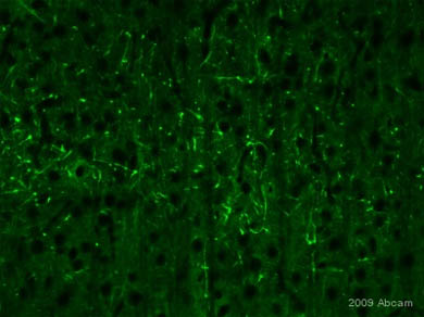 ab37500 staining NUMBL in rat brain tissue section by Immunohistochemistry (PFA perfusion fixed frozen sections). Tissue from 4% PFA perfused animals underwent overnight fixation in 4% paraformaldehyde, cryoprotected in 30% sucrose and cut using cryostat.The primary antibody was diluted, 1/1000 (PBS + 0.3% Triton X100) and incubated with sample for 18 hours at 20°C. An Alexa Fluor® 488 conjugated goat polyclonal to rabbit IgG was used undiluted as secondary.
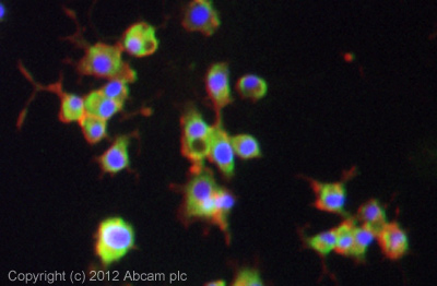 ICC/IF image of ab37500 stained pc12 cells. The cells were 4% formaldehyde fixed (10 min) and then incubated in 1%BSA / 10% normal goat serum / 0.3M glycine in 0.1% PBS-Tween for 1h to permeabilise the cells and block non-specific protein-protein interactions. The cells were then incubated with the antibody (ab37500, 1µg/ml) overnight at +4°C. The secondary antibody (green) was ab96899, DyLight® 488 goat anti-rabbit IgG (H+L) used at a 1/250 dilution for 1h. Alexa Fluor® 594 WGA was used to label plasma membranes (red) at a 1/200 dilution for 1h. DAPI was used to stain the cell nuclei (blue) at a concentration of 1.43µM.
Product References
Numbl inhibits glioma cell migration and invasion by suppressing TRAF5-mediated - Numbl inhibits glioma cell migration and invasion by suppressing TRAF5-mediated
Tao T, Cheng C, Ji Y, Xu G, Zhang J, Zhang L, Shen A. Mol Biol Cell. 2012 Jul;23(14):2635-44.
Epigenetic regulation of miR-184 by MBD1 governs neural stem cell proliferation - Epigenetic regulation of miR-184 by MBD1 governs neural stem cell proliferation
Liu C, Teng ZQ, Santistevan NJ, Szulwach KE, Guo W, Jin P, Zhao X. Cell Stem Cell. 2010 May 7;6(5):433-44.
NUMBL interacts with TRAF6 and promotes the degradation of TRAF6. - NUMBL interacts with TRAF6 and promotes the degradation of TRAF6.
Zhou L, Ma Q, Shi H, Huo K. Biochem Biophys Res Commun. 2010 Feb 12;392(3):409-14. doi: