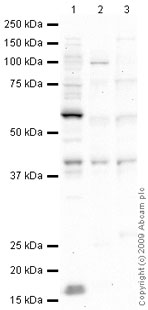
All lanes : Anti-p53R2 antibody (ab63814) at 1 µg/mlLane 1 : MCF7 (Human breast adenocarcinoma cell line) Whole Cell LysateLane 2 : Skeletal Muscle (Human) Tissue Lysate - adult normal tissue (ab29330)Lane 3 : Placenta (Human) Tissue Lysate - adult normal tissue (ab29745)Lysates/proteins at 10 µg per lane.SecondaryGoat polyclonal to Rabbit IgG - H&L - Pre-Adsorbed (HRP) at 1/3000 dilutionPerformed under reducing conditions.
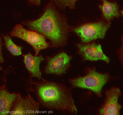
ICC/IF image of ab63814 stained HeLa cells. The cells were 4% PFA fixed (10 min) and then incubated in 1%BSA / 10% normal Goat serum / 0.3M glycine in 0.1% PBS-Tween for 1h to permeabilise the cells and block non-specific protein-protein interactions. The cells were then incubated with the antibody (ab63814, 1µg/ml) overnight at +4°C. The secondary antibody (green) was Alexa Fluor® 488 Goat anti-Rabbit IgG (H+L) used at a 1/1000 dilution for 1h. Alexa Fluor® 594 WGA was used to label plasma membranes (red) at a 1/200 dilution for 1h. DAPI was used to stain the cell nuclei (blue). This antibody also gave a positive result in 4% PFA fixed (10 min) MCF cells at 1µg/ml.
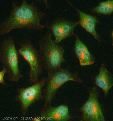
ICC/IF image of ab63814 stained HeLa cells. The cells were 4% pfa fixed (10 min) and then incubated in 1%BSA / 10% normal Goat serum / 0.3M glycine in 0.1% PBS-Tween for 1h to permeabilise the cells and block non-specific protein-protein interactions. The cells were then incubated with the antibody (ab63814, 1µg/ml) overnight at +4°C. The secondary antibody (green) was Alexa Fluor® 488 Goat anti-Rabbit IgG (H+L) used at a 1/1000 dilution for 1h. Alexa Fluor® 594 WGA was used to label plasma membranes (red) at a 1/200 dilution for 1h. DAPI was used to stain the cell nuclei (blue). This antibody also gave a positive result in 4% pfa fixed (10 min) HepG2, MCF-7 cells at 1µg/ml.