
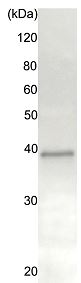
Anti-Rad51 antibody - ChIP Grade (ab176458) at 1/1000 dilution + Crude HeLa cell extracts at 10 µgSecondaryGoat anti-Rabbit IgG conjugated to HRP at 1/20000 dilution
ab176458 (20 µg) was incubated with 20 μg of HeLa cell extract, and precipitated with 20 μg of proteinA-beads. The sample was dissociated from the precipitate by heating in SDS-sample buffer and analyzed by western blotting with anti-Rad51 antiserum (chicken, ab63802) at 1/1000 dilution. As secondary antibody, anti-chicken IgG antibody (rabbit) was used at 1/10000 dilution.
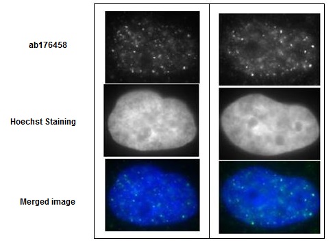
Immunofluorescence detection of Rad51 foci formation after X-ray irradiation in GM0637 cells with ab176458 at 1/10000 dilution (left panels) and 1/1000 dilution (right panels). The secondary antibody, anti-rabbit Alexa 488 was used at 1/10000 dilution. Cells were irradiated by X-rays at 2 Gy, grown for 1 hr, fixed with 4% paraformaldehyde in 1x PBS for 10 min, washed 3 times with PBS for 3 min, permealized by treatment with 0.5% Triton for 5 min, washed 3 times with PBS for 3 min, incubated with ab176458 for 30 min at 37°C, washed 3 times with PBS for 3 min, incubated with secondary antibody for 30 min at 37°C, washed 3 times with PBS for 3 min, stained with Hoechst for 1 min and mounted. The pictures were by courtesy of Prof. S. Tashiro and Dr. K. Kono at Hiroshima University.
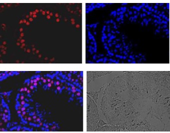
Immunohistological staining of Rad51 protein in mouse testis using ab176458. A section of formalin fixed and paraffin embedded mouse testis was treated with ab176458 at 1/100 dilution after deparaffization and antigen retrieval. The secondary antibody, Alexa Fluor® 647conjugated anti-rabbit IgG was used at 1/1,000 dilution (top left). The sample was counter-stained with DAPI (top right) and the merged image is shown (bottom left). The white light image of the same region is shown on the bottom right.