![All lanes : Anti-SFRS3 antibody [EPR16976] (ab198291) at 1/5000 dilutionLane 1 : Jurkat (Human T cell leukemia cells from peripheral blood) whole cell lysateLane 2 : HepG2 (Human liver hepatocellular carcinoma) whole cell lysateLane 3 : K562 (Human chronic myelogenous leukemia cells from bone marrow) whole cell lysateLysates/proteins at 20 µg per lane.SecondaryGoat Anti-Rabbit IgG, (H+L), Peroxidase conjugated at 1/1000 dilution](http://www.bioprodhub.com/system/product_images/ab_products/2/sub_4/29186_ab198291-240236-198291.JPG)
All lanes : Anti-SFRS3 antibody [EPR16976] (ab198291) at 1/5000 dilutionLane 1 : Jurkat (Human T cell leukemia cells from peripheral blood) whole cell lysateLane 2 : HepG2 (Human liver hepatocellular carcinoma) whole cell lysateLane 3 : K562 (Human chronic myelogenous leukemia cells from bone marrow) whole cell lysateLysates/proteins at 20 µg per lane.SecondaryGoat Anti-Rabbit IgG, (H+L), Peroxidase conjugated at 1/1000 dilution
![All lanes : Anti-SFRS3 antibody [EPR16976] (ab198291) at 1/1000 dilutionLane 1 : Mouse brain lysateLane 2 : Rat brain lysateLane 3 : C6 (Rat glial tumor) whole cell lysateLane 4 : RAW 264.7(Mouse macrophage cells transformed with Abelson murine leukemia virus) whole cell lysateLane 5 : PC-12 (Rat adrenal gland pheochromocytoma) whole cell lysateLane 6 : NIH/3T3 (Mouse embryo fibroblast cells) whole cell lysateLysates/proteins at 10 µg per lane.SecondaryGoat Anti-Rabbit IgG, (H+L), Peroxidase conjugated at 1/1000 dilution](http://www.bioprodhub.com/system/product_images/ab_products/2/sub_4/29187_ab198291-240235-1982912.JPG)
All lanes : Anti-SFRS3 antibody [EPR16976] (ab198291) at 1/1000 dilutionLane 1 : Mouse brain lysateLane 2 : Rat brain lysateLane 3 : C6 (Rat glial tumor) whole cell lysateLane 4 : RAW 264.7(Mouse macrophage cells transformed with Abelson murine leukemia virus) whole cell lysateLane 5 : PC-12 (Rat adrenal gland pheochromocytoma) whole cell lysateLane 6 : NIH/3T3 (Mouse embryo fibroblast cells) whole cell lysateLysates/proteins at 10 µg per lane.SecondaryGoat Anti-Rabbit IgG, (H+L), Peroxidase conjugated at 1/1000 dilution
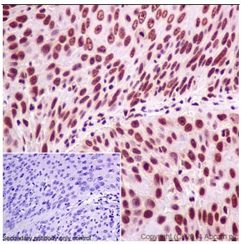
Immunohistochemical analysis of paraffin-embedded Human cervix carcinoma tissue labeling SFRS3 with ab198291 at 1/500 dilution followed by Goat Anti-Rabbit IgG H&L (HRP) (ab97051) at 1/500 dilution. Nuclear staining on Human cervix carcinoma tissue is observed. Counter stained with Hematoxylin.Negative control: Used PBS instead of primary ab.
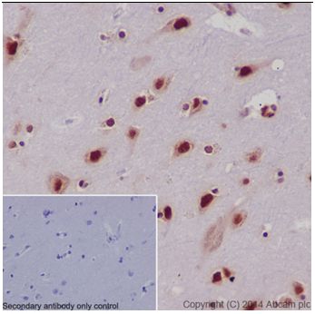
Immunohistochemical analysis of paraffin-embedded Human cerebral cortex tissue labeling SFRS3 with ab198291 at 1/500 dilution followed by Goat Anti-Rabbit IgG H&L (HRP) (ab97051) at 1/500 dilution. Nuclear and weakly cytoplasm staining on Human cerebral cortex tissue is observed. Counter stained with Hematoxylin.Negative control: Used PBS instead of primary ab.
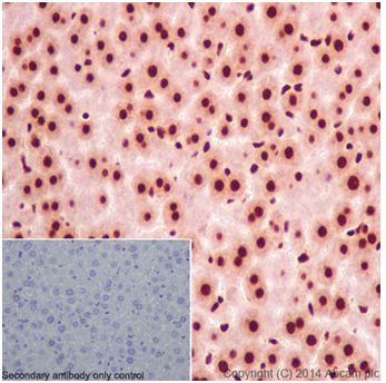
Immunohistochemical analysis of paraffin-embedded Mouse liver tissue labeling SFRS3 with ab198291 at 1/500 dilution followed by Goat Anti-Rabbit IgG H&L (HRP) (ab97051) at 1/500 dilution. Nuclear and weakly cytoplasm staining on mouse liver tissue is observed. Counter stained with Hematoxylin.Negative control: Used PBS instead of primary ab.

Flow cytometric analysis of 2% paraformaldehyde-fixed Jurkat (Human T Cell Leukemia cells from peripheral blood) labeling SFRS3 with ab198291 at 1/440 dilution (red) compared with a Rabbit monoclonal IgG isotype control (black) and an unlabelled control (cells without incubation with primary antibody and secondary antibody; blue). Goat anti rabbit IgG (FITC) at 1/150 dilution was used as the secondary antibody.