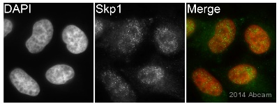
ab124473 staining Skp1 in HeLa cells by ICC/IF (Immunocytochemistry/immunofluorescence). Cells were fixed with paraformaldehyde and permeabilized with 0.5% Triton X-100 in PBS. Samples were incubated with primary antibody (1/200 in PBS) for 16 hours at 22°C. An Alexa Fluor® 488-conjugated goat anti-mouse IgG polyclonal (1/200) was used as the secondary antibody.See Abreview
![All lanes : Anti-Skp1 antibody [1H9] (ab124473) at 1/500 dilutionLane 1 : HeLa cell lysateLane 2 : Raji cell lysateLane 3 : Jurkat cell lysateLane 4 : MCF7 cell lysateLane 5 : HepG2 cell lysateLane 6 : PC12 cell lysateLane 7 : COS7 cell lysate](http://www.bioprodhub.com/system/product_images/ab_products/2/sub_5/471_Skp1-Primary-antibodies-ab124473-1.jpg)
All lanes : Anti-Skp1 antibody [1H9] (ab124473) at 1/500 dilutionLane 1 : HeLa cell lysateLane 2 : Raji cell lysateLane 3 : Jurkat cell lysateLane 4 : MCF7 cell lysateLane 5 : HepG2 cell lysateLane 6 : PC12 cell lysateLane 7 : COS7 cell lysate
![Anti-Skp1 antibody [1H9] (ab124473) at 1/500 dilution + Human Skp1 recombinant protein (aa: 1-160)](http://www.bioprodhub.com/system/product_images/ab_products/2/sub_5/472_Skp1-Primary-antibodies-ab124473-2.jpg)
Anti-Skp1 antibody [1H9] (ab124473) at 1/500 dilution + Human Skp1 recombinant protein (aa: 1-160)
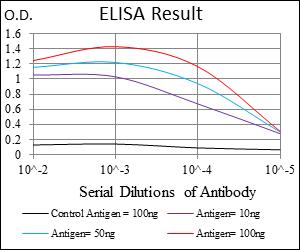
ELISA using ab124473.

ab124473, at 1/200 dilution, staining Skp1 in paraffin-embedded Human ovarian cancer tissue by immunohistochemistry followed by DAB staining.
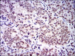
ab124473, at 1/200 dilution, staining Skp1 in paraffin-embedded Human cervical cancer tissue by immunohistochemistry followed by DAB staining.
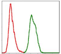
ab124473, at 1/200 dilution, staining Skp1 in HeLa cells by flow cytometry (green) or negative control (red).