![Anti-SUNC1 antibody [EPR12910] (ab181063) at 1/1000 dilution + LnCap cell lysate at 20 µgSecondaryGoat Anti-Rabbit IgG, (H+L), Peroxidase conjugated at 1/1000 dilution](http://www.bioprodhub.com/system/product_images/ab_products/2/sub_5/6856_ab181063-213199-ab1810631.jpg)
Anti-SUNC1 antibody [EPR12910] (ab181063) at 1/1000 dilution + LnCap cell lysate at 20 µgSecondaryGoat Anti-Rabbit IgG, (H+L), Peroxidase conjugated at 1/1000 dilution
![Anti-SUNC1 antibody [EPR12910] (ab181063) at 1/1000 dilution + HepG2 cell lysate at 10 µgSecondaryGoat Anti-Rabbit IgG, (H+L), Peroxidase conjugated at 1/1000 dilution](http://www.bioprodhub.com/system/product_images/ab_products/2/sub_5/6857_ab181063-213198-ab1810632.jpg)
Anti-SUNC1 antibody [EPR12910] (ab181063) at 1/1000 dilution + HepG2 cell lysate at 10 µgSecondaryGoat Anti-Rabbit IgG, (H+L), Peroxidase conjugated at 1/1000 dilution
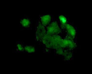
Immunofluorescent analysis of paraformaldehyde-fixed HepG2 cells labeling SUNC1 with ab181063 at 1/100 dilution. A Goat anti rabbit IgG (Alexa Fluor®488) was used as the secondary at 1/200 dilution.
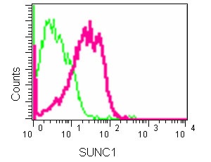
Flow Cytometric analysis of HepG2 cells (2% paraformaldehyde-fixed) labeling SUNC1 with ab181063 at 1/110 dilution (red) or a rabbit IgG (negative) (green), followed by Goat anti rabbit IgG (FITC) secondary at 1/150 dilution.