Anti-CDIPT Antibody
| Name | Anti-CDIPT Antibody |
|---|---|
| Supplier | LifeSpan Bioscience |
| Catalog | LS-C168611 |
| Prices | $295.00 |
| Sizes | 400 µl |
| Host | Rabbit |
| Clonality | Polyclonal |
| Applications | IHC-P WB FC |
| Species Reactivities | Human, Mouse |
| Antigen | CDIPT antibody was raised against kLH-conjugated synthetic peptide selected from internal region of human CDIPT. |
| Purity/Format | Immunoaffinity purified |
| Blocking Peptide | CDH7 / Cadherin 7 Antibody Blocking Peptide |
| Description | Rabbit Polyclonal |
| Gene | CDIPT |
| Supplier Page | Shop |
Product images
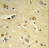 Formalin-fixed and paraffin-embedded human brain tissue reacted with CDIPT Antibody , which was peroxidase-conjugated to the secondary antibody, followed by DAB staining. This data demonstrates the use of this antibody for immunohistochemistry; clinical relevance has not been evaluated.
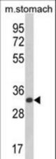 Western blot of CDIPT Antibody in mouse stomach tissue lysates (35 ug/lane). CDIPT (arrow) was detected using the purified antibody.
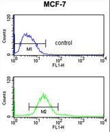 CDIPT Antibody flow cytometry of MCF-7 cells (bottom histogram) compared to a negative control cell (top histogram). FITC-conjugated goat-anti-rabbit secondary antibodies were used for the analysis.