Anti-HCLS1 Antibody (aa362-391)
| Name | Anti-HCLS1 Antibody (aa362-391) |
|---|---|
| Supplier | LifeSpan Bioscience |
| Catalog | LS-C166815 |
| Prices | $295.00 |
| Sizes | 400 µl |
| Host | Rabbit |
| Clonality | Polyclonal |
| Applications | IHC-P WB ELISA |
| Species Reactivities | Human |
| Purity/Format | Protein A purified |
| Blocking Peptide | HCK Antibody Blocking Peptide |
| Description | Rabbit Polyclonal |
| Gene | HCLS1 |
| Conjugate | Unconjugated |
| Supplier Page | Shop |
Product images
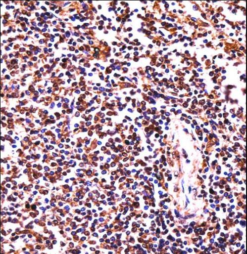 HCLS1 Antibody immunohistochemistry of formalin-fixed and paraffin-embedded human spleen tissue followed by peroxidase-conjugated secondary antibody and DAB staining.
 Western blot of lysates from K562, Raji cell line (from left to right), using HCLS1 Antibody. Antibody was diluted at 1:1000 at each lane. A goat anti-rabbit IgG H&L (HRP) at 1:5000 dilution was used as the secondary antibody. Lysates at 35ug per lane.
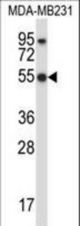 HCLS1 Antibody western blot of MDA-MB231 cell line lysates (35 ug/lane). The HCLS1 antibody detected the HCLS1 protein (arrow).