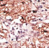
Formalin-fixed and paraffin-embedded human cancer tissue reacted with the primary antibody, which was peroxidase-conjugated to the secondary antibody, followed by DAB staining. This data demonstrates the use of this antibody for immunohistochemistry; clinical relevance has not been evaluated. BC = breast carcinoma; HC = hepatocarcinoma.
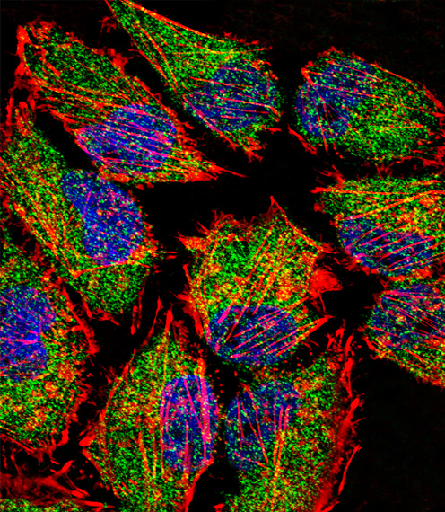
Fluorescent confocal image of HeLa cell stained with LSD1 Antibody. HeLa cells were fixed with 4% PFA (20 min), permeabilized with Triton X-100 (0.1%, 10 min), then incubated with LSD1 primary antibody (1:25, 1 h at 37°C). For secondary antibody, Alexa Fluor 488 conjugated donkey anti-rabbit antibody (green) was used (1:400, 50 min at 37°C). Cytoplasmic actin was counterstained with Alexa Fluor 555 (red) conjugated Phalloidin (7units/ml, 1 h at 37°C). Nuclei were counterstained with DAPI (blue) (10 ug/ml, 10 min). LSD1 immunoreactivity is localized to Cytoplasm significantly and Nucleus weakly.

Western blot of anti-LSD1 antibody in mouse brain tissue lysate (35 ug/lane). LSD1(arrow) was detected using the purified antibody.
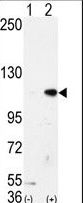
Western blot of AOF2 (arrow) using LSD1 Antibody. 293 cell lysates (2 ug/lane) either nontransfected (Lane 1) or transiently transfected with the AOF2 gene (Lane 2) (Origene Technologies).