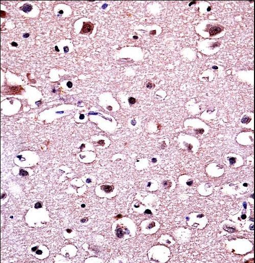
Parkin Antibody immunohistochemistry of formalin-fixed and paraffin-embedded human brain tissue followed by peroxidase-conjugated secondary antibody and DAB staining.
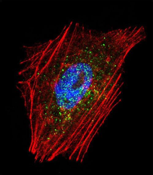
Confocal immunofluorescent of Parkin Antibody with NCI-H460 cell followed by Alexa Fluor 488-conjugated goat anti-rabbit lgG (green). Actin filaments have been labeled with Alexa Fluor 555 phalloidin (red). DAPI was used to stain the cell nuclear (blue).
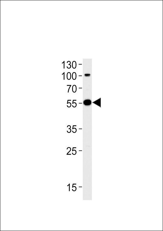
Western blot of lysate from SH-SY5Y cell line, using Park2 Antibody. Antibody was diluted at 1:1000 at each lane. A goat anti-rabbit (HRP) at 1:5000 dilution was used as the secondary antibody. Lysate at 35ug.
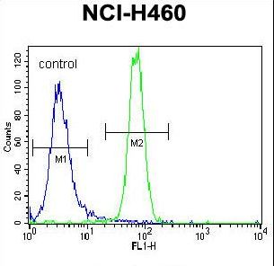
Parkin Antibody flow cytometry of NCI-H460 cells (right histogram) compared to a negative control cell (left histogram). FITC-conjugated goat-anti-rabbit secondary antibodies were used for the analysis.