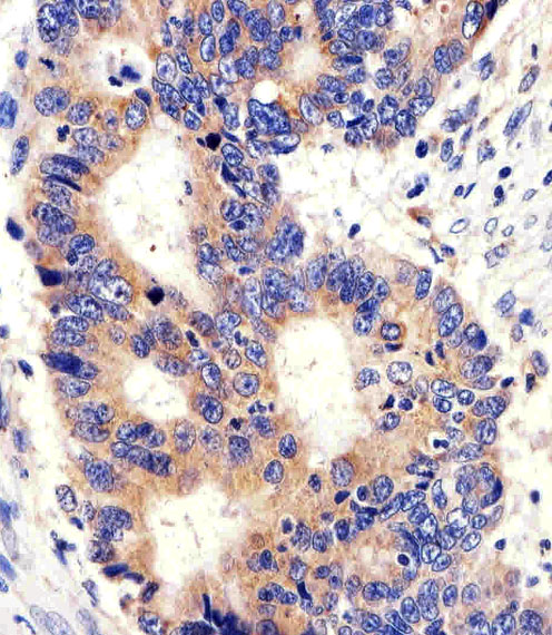
Immunohistochemical of paraffin-embedded H.colon carcinoma section using PCSK9 Antibody. Antibody was diluted at 1:100 dilution. A peroxidase-conjugated goat anti-rabbit IgG at 1:400 dilution was used as the secondary antibody, followed by DAB staining.
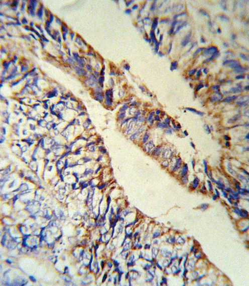
PCSK9 Antibody (RB18880) IHC of formalin-fixed and paraffin-embedded human Colon carcinoma followed by peroxidase-conjugated secondary antibody and DAB staining.
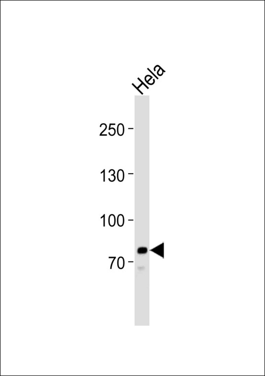
Western blot of lysate from HeLa cell line, using PCSK9 Antibody. Antibody was diluted at 1:1000. A goat anti-rabbit IgG H&L (HRP) at 1:10000 dilution was used as the secondary antibody. Lysate at 35ug.
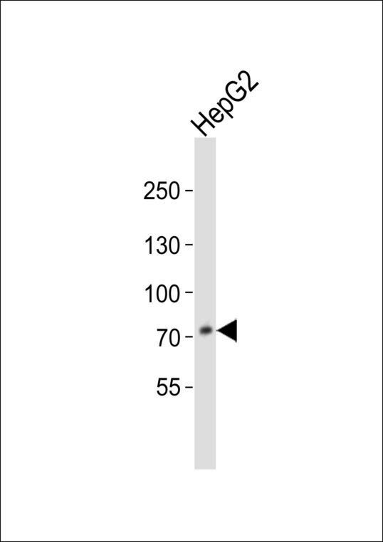
Western blot of lysate from HepG2 cell line, using PCSK9 Antibody. Antibody was diluted at 1:1000. A goat anti-rabbit IgG H&L (HRP) at 1:10000 dilution was used as the secondary antibody. Lysate at 20ug.
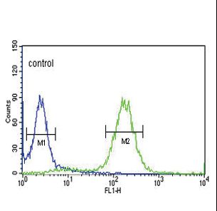
PCSK9 Antibody flow cytometry of HeLa cells (right histogram) compared to a negative control cell (left histogram). FITC-conjugated goat-anti-rabbit secondary antibodies were used for the analysis.