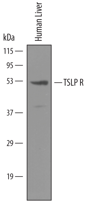
Western blot shows lysates of human liver tissue. PVDF membrane was probed with 1 µg/mL of Goat Anti-Human TSLP R Antigen Affinity-purified Polyclonal Antibody (Catalog # AF981) followed by HRP-conjugated Anti-Goat IgG Secondary Antibody (Catalog # HAF019). A specific band was detected for TSLP R at approximately 50 kDa (as indicated). This experiment was conducted under reducing conditions and using Immunoblot Buffer Group 8.
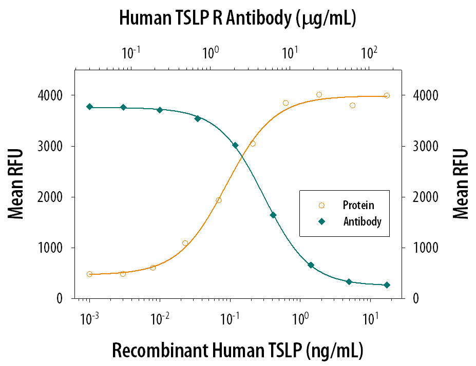
Recombinant Human TSLP (Catalog # 1398-TS) stimulates proliferation in the BaF3 mouse pro‑B cell line co-transfected with human IL‑7 R alpha and TSLP R in a dose-dependent manner (orange line). Proliferation elicited by Recombinant Human TSLP (0.3 ng/mL) is neutralized (green line) by increasing concentrations of Goat Anti-Human TSLP R Antigen Affinity-purified Polyclonal Antibody (Catalog # AF981). The ND50 is typically 1‑5 µg/mL.
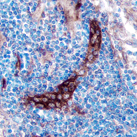
TSLP R was detected in immersion fixed paraffin-embedded sections of human lymph node using Goat Anti-Human TSLP R Antigen Affinity-purified Polyclonal Antibody (Catalog # AF981) at 5 µg/mL overnight at 4 °C. Tissue was stained using the Anti-Goat HRP-DAB Cell & Tissue Staining Kit (brown; Catalog # CTS008) and counterstained with hematoxylin (blue). Specific staining was localized to the plasma membrane. View our protocol for Chromogenic IHC Staining of Paraffin-embedded Tissue Sections.
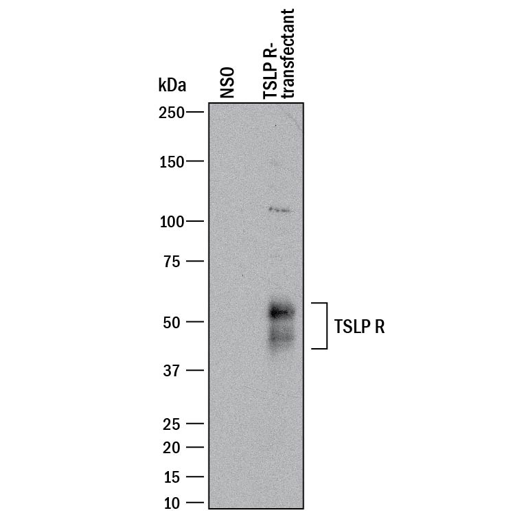
Western blot shows lysates of NS0 mouse myeloma cell line either mock transfected or transfected with human TSLP R. PVDF membrane was probed with 0.2 µg/mL of Goat Anti-Human TSLP R Antigen Affinity-purified Polyclonal Antibody (Catalog # AF981) followed by HRP-conjugated Anti-Goat IgG Secondary Antibody (Catalog # HAF017). Specific bands were detected for TSLP R at approximately 45-60 kDa (as indicated). This experiment was conducted under reducing conditions and using Immunoblot Buffer Group 1.