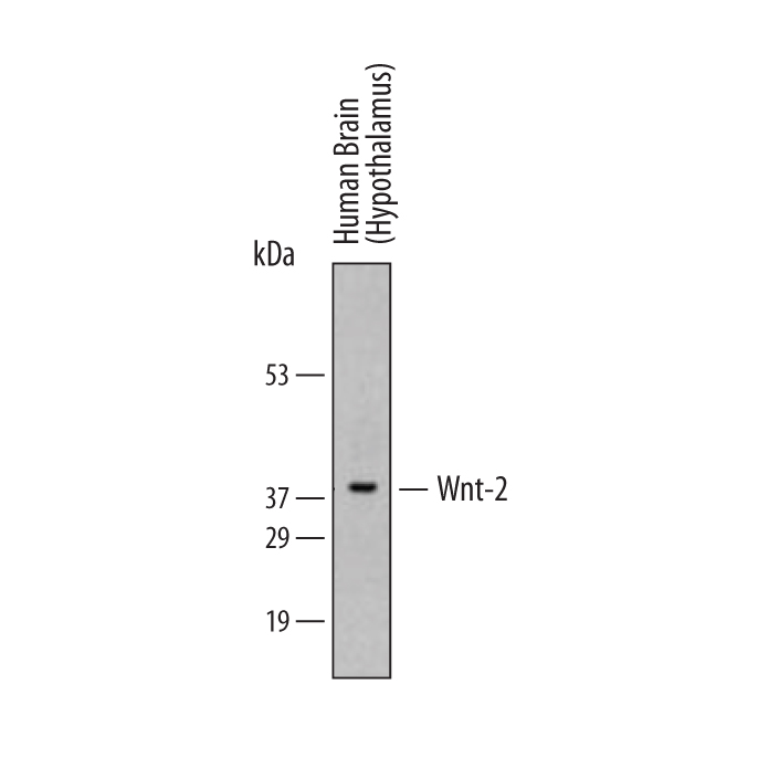
Western blot shows lysates of human brain (hypothalamus) tissue. PVDF membrane was probed with 1 µg/mL of Goat Anti-Human Wnt‑2 Antigen Affinity-purified Polyclonal Antibody (Catalog # AF3464) followed by HRP-conjugated Anti-Goat IgG Secondary Antibody (Catalog # HAF019). A specific band was detected for Wnt‑2 at approximately 40 kDa (as indicated). This experiment was conducted under reducing conditions and using Immunoblot Buffer Group 2.

Wnt‑2 was detected in immersion fixed paraffin-embedded sections of human stomach cancer tissue using 10 µg/mL Goat Anti-Human Wnt‑2 Antigen Affinity-purified Polyclonal Antibody (Catalog # AF3464) overnight at 4 °C. Before incubation with the primary antibody tissue was subjected to heat-induced epitope retrieval using Antigen Retrieval Reagent-Basic (Catalog # CTS013). Tissue was stained with the Anti-Goat HRP-DAB Cell & Tissue Staining Kit (brown; Catalog # CTS008) and counterstained with hematoxylin (blue). View our protocol for Chromogenic IHC Staining of Paraffin-embedded Tissue Sections.