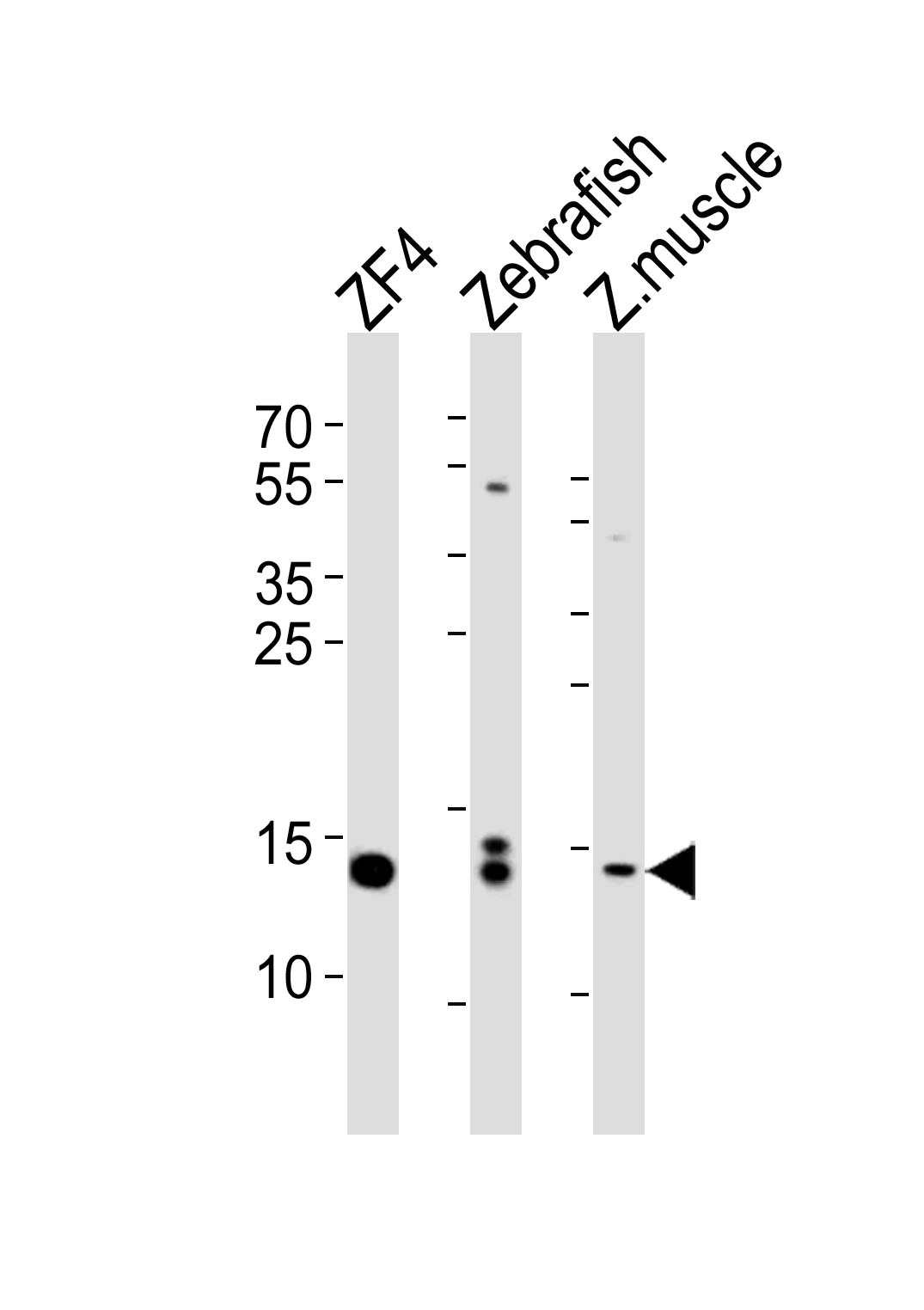
Western blot analysis of lysates from ZF4 cell line, Zebrafish, zebra fish muscle tissue lysate(from left to right), using HIST1H2BJ Antibody (Center)(Cat. #AP10727c). AP10727c was diluted at 1:1000 at each lane. A goat anti-rabbit IgG H&L(HRP) at 1:5000 dilution was used as the secondary antibody. Lysates at 35ug per lane.
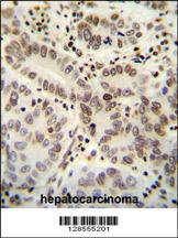
HIST1H2B antibody (Center) (Cat. #AP10727c) immunohistochemistry analysis in formalin fixed and paraffin embedded human hepatocarcinoma followed by peroxidase conjugation of the secondary antibody and DAB staining. This data demonstrates the use of the HIST1H2B antibody (Center) for immunohistochemistry. Clinical relevance has not been evaluated.
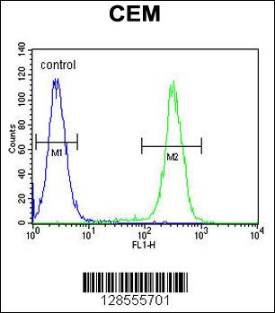
HIST1H2B Antibody (Center) (Cat. #AP10727c) flow cytometric analysis of CEM cells (right histogram) compared to a negative control cell (left histogram).FITC-conjugated goat-anti-rabbit secondary antibodies were used for the analysis.
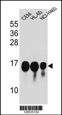
HIST1H2B Antibody (Center) (Cat. #AP10727c) western blot analysis in CEM,HL-60,NCI-H460 cell line lysates (35ug/lane).This demonstrates the HIST1H2B antibody detected the HIST1H2B protein (arrow).