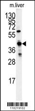
Western blot analysis of GCAT antibody (Center) (Cat.#AP7498c) in mouse liver tissue lysates (35ug/lane). GCAT (arrow) was detected using the purified Pab.
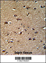
Formalin-fixed and paraffin-embedded human brain reacted with GCAT Antibody (Center), which was peroxidase-conjugated to the secondary antibody, followed by DAB staining. This data demonstrates the use of this antibody for immunohistochemistry; clinical relevance has not been evaluated.
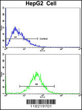
Flow cytometric analysis of HepG2 cells using GCAT Antibody (Center)(bottom histogram) compared to a negative control cell (top histogram). FITC-conjugated goat-anti-rabbit secondary antibodies were used for the analysis.
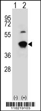
Western blot analysis of GCAT (arrow) using rabbit polyclonal GCAT Antibody (Center) (Cat.#AP7498c). 293 cell lysates (2 ug/lane) either nontransfected (Lane 1) or transiently transfected (Lane 2) with the GCAT gene.