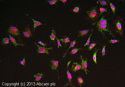
ab128911 stained HepG2 cells. The cells were 4% formaldehyde fixed for 10 minutes at room temperature and then incubated in 1%BSA / 10% normal goat serum / 0.3M glycine in 0.1% PBS-Tween for 1hour at room temperature to permeabilise the cells and block non-specific protein-protein interactions. The cells were then incubated with the antibody (ab128911 at 1/100 dilution) overnight at +4°C. The secondary antibody (pseudo-colored green) was Goat Anti-Rabbit IgG H&L (Alexa Fluor® 488) preadsorbed (ab150081) used at a 1/1000 dilution for 1hour at room temperature. Alexa Fluor® 594 WGA was used to label plasma membranes (pseudo-colored red) at a 1/200 dilution for 1hour at room temperature. DAPI was used to stain the cell nuclei (pseudo-colored blue) at a concentration of 1.43µM for 1hour at room temperature.
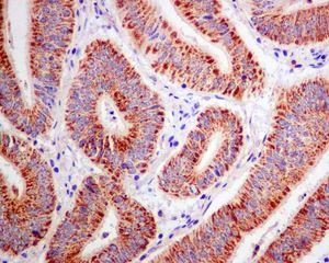
ab128911 at 1/100 dilution staining ACAA2 in paraffin embedded Human colonic carcinoma tissue by immunohistochemistry.
![All lanes : Anti-ACAA2 antibody [EPR6733] (ab128911) at 1/1000 dilutionLane 1 : HepG2 cell lysateLane 2 : 293T cell lysateLane 3 : HeLa cell lysateLysates/proteins at 10 µg per lane.SecondaryHRP labelled goat anti-rabbit at 1/2000 dilution](http://www.bioprodhub.com/system/product_images/ab_products/2/sub_1/1423_ACAA2-Primary-antibodies-ab128911-1.jpg)
All lanes : Anti-ACAA2 antibody [EPR6733] (ab128911) at 1/1000 dilutionLane 1 : HepG2 cell lysateLane 2 : 293T cell lysateLane 3 : HeLa cell lysateLysates/proteins at 10 µg per lane.SecondaryHRP labelled goat anti-rabbit at 1/2000 dilution
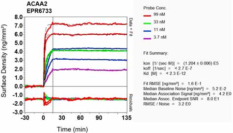
Equilibrium disassociation constant (KD)Learn more about KD Click here to learn more about KD