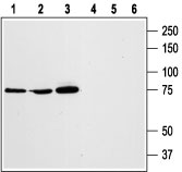
Western blot analysis of RBL (lanes 1 and 4), HL-60 (lanes 2 and 5), and Jurkat (lanes 3 and 6) cell lysates: 1, 2, 3. Anti-STIM1 (extracellular) antibody (#AG1335), (1:1000). 4, 5, 6. Anti-STIM1 (extracellular) antibody, preincubated with the control peptide antigen.
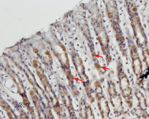
Expression of STIM1 in rat stomach Immunohistochemical staining of paraffin embedded rat stomach sections using Anti-STIM1 (extracellular) antibody (#AG1335), (1:100). STIM1 is expressed in the parietal cells of the gastric mucosa (arrows). Hematoxilin is used as the counterstain.
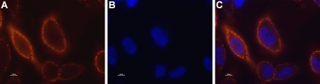
Expression of STIM1 in RBL cells Immunocytochemical staining of STIM1 in live rat basophilic leukemia (RBL) cells. A. Extracellular staining of cells with Anti-STIM1 (extracellular) antibody (#AG1335), (1:50) followed by goat anti-rabbit-AlexaFluor-555 secondary antibody. B. Nuclear staining of cells using the cell-permeable dye Hoechst 33342. C. Merged image of panels A and B.
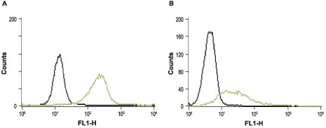
Indirect flow cytometry analysis of intact RBL (A) and Jurkat (B) cells. ___ Cells + FITC-conjugated goat anti-rabbit antibody.___ Cells + Anti-STIM1 (extracellular) antibody (#AG1335), (5-10 µg antibody/0.5-1x106 cells) + FITC-conjugated goat anti-rabbit antibody.