![Anti-AIF antibody [7F7AB10] (ab110327) at 5 µg/ml + Isolated mitochondria from Human heart at 5 µg](http://www.bioprodhub.com/system/product_images/ab_products/2/sub_1/3840_AIF-Primary-antibodies-ab110327-1.jpg)
Anti-AIF antibody [7F7AB10] (ab110327) at 5 µg/ml + Isolated mitochondria from Human heart at 5 µg
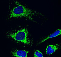
Immunocytochemistry analysis using ab110327 at 5µg/ml staining AIF in HeLa cells (4% paraformaldehyde fixed and Triton X-100 permeabilized). The secondary antibody was (green) Alexa Fluor® 488 goat anti-mouse IgG (H+L) used at a 1/1000 dilution for 1 hour. DAPI was used to stain the cell nuclei (blue). Target protein locates mainly in mitochondria.

Flow cytometric analysis using ab110327 at 1µg/ml staining AIF in HL60 cells (blue). Isotype control antibody (red).
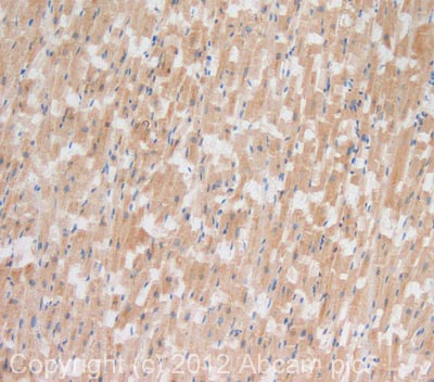
IHC image of AIF staining in human heart formalin fixed paraffin embedded tissue section, performed on a Leica BondTM system using the standard protocol F. The section was pre-treated using heat mediated antigen retrieval with sodium citrate buffer (pH6, epitope retrieval solution 1) for 20 mins. The section was then incubated with ab110327, 10µg/ml, for 15 mins at room temperature and detected using an HRP conjugated compact polymer system. DAB was used as the chromogen. The section was then counterstained with haematoxylin and mounted with DPX. For other IHC staining systems (automated and non-automated) customers should optimize variable parameters such as antigen retrieval conditions, primary antibody concentration and antibody incubation times.