![Anti-Apolipoprotein CI antibody [EPR16813] (ab198288) at 1/1000 dilution + Human fetal liver lysate at 20 µgSecondaryAnti-Rabbit IgG (HRP), specific to the non-reduced form of IgG at 1/1000 dilution](http://www.bioprodhub.com/system/product_images/ab_products/2/sub_1/8460_ab198288-240160-198288.JPG)
Anti-Apolipoprotein CI antibody [EPR16813] (ab198288) at 1/1000 dilution + Human fetal liver lysate at 20 µgSecondaryAnti-Rabbit IgG (HRP), specific to the non-reduced form of IgG at 1/1000 dilution
![Anti-Apolipoprotein CI antibody [EPR16813] (ab198288) at 1/5000 dilution + Human plasma lysate at 20 µgSecondaryAnti-Rabbit IgG (HRP), specific to the non-reduced form of IgG at 1/1000 dilution](http://www.bioprodhub.com/system/product_images/ab_products/2/sub_1/8461_ab198288-240159-1982882.JPG)
Anti-Apolipoprotein CI antibody [EPR16813] (ab198288) at 1/5000 dilution + Human plasma lysate at 20 µgSecondaryAnti-Rabbit IgG (HRP), specific to the non-reduced form of IgG at 1/1000 dilution
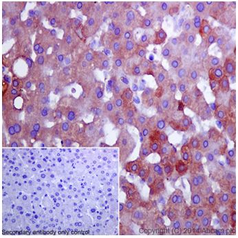
Immunohistochemical analysis of paraffin-embedded Human liver tissue labeling Apolipoprotein CI with ab198288 at 1/1600 dilution followed by Goat Anti-Rabbit IgG H&L (HRP) (ab97051) at 1/500 dilution. Cytoplasm staining on Human liver tissue is observed. Counter stained with Hematoxylin.Negative control: Used PBS instead of primary ab.
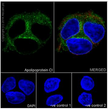
Immunofluorescent analysis of 4% paraformaldehyde-fixed, 0.1% Triton X-100 permeabilized HepG2 (Human liver hepatocellular carcinoma) cells labeling Apolipoprotein CI with ab198288 at 1/800 dilution, followed by Goat anti-rabbit IgG (Alexa Fluor® 488) (ab150077) secondary antibody at 1/500 dilution (green). Cytoplasm staining on HepG2 cell line is observed. The nuclear counterstain is DAPI (blue). Tubulin is detected with ab7291 (anti-Tubulin mouse mAb) at 1/1000 dilution and ab150120 (AlexaFluor®594 Goat anti-Mouse secondary) at 1/500 dilution (red).The negative controls are as follows:--ve control 1 - ab198288 at 1/800 dilution followed by ab150120 (AlexaFluor®594 Goat anti-Mouse secondary) at 1/500 dilution.-ve control 2. - ab7291 (anti-Tubulin mouse mAb) at 1/1000 dilution followed by ab150077 (Alexa Fluor®488 Goat Anti-Rabbit IgG H&L) at 1/500 dilution.
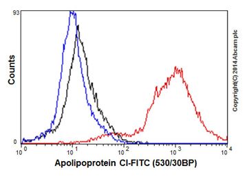
Flow cytometric analysis of 2% paraformaldehyde-fixed HeLa (Human epithelial cells from cervix adenocarcinoma) cells labeling Apolipoprotein CI with ab198288 at 1/80 dilution (red). The secondary antibody was Goat anti rabbit IgG (FITC) at 1/150 dilution. The isotype control is Rabbit monoclonal IgG (black) (ab172730) and cell without incubation with primary antibody and secondary antibody is blue.
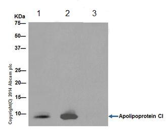
Immunoprecipitation analysis of Human plasma whole cell extract labeling Apolipoprotein CI using ab198288 at 1/40 dilution (Lane 2).Lane 3: IP using Rabbit monoclonal IgG (ab172730) instead of ab198288 in Human plasma whole cell extract.Lane 1: Input: 10 μg Human plasma whole cell extract.Subsequent WB detection was performed using ab198288 at 1/1000 dilution.An Anti-Rabbit IgG (HRP), specific to the non-reduced form of IgG at 1/1500 was used as secondary antibody.