Human Cadherin-11 MAb (Clone 283416), Mouse IgG2B
| Name | Human Cadherin-11 MAb (Clone 283416), Mouse IgG2B |
|---|---|
| Supplier | R&D Systems |
| Catalog | MAB1790-SP |
| Host | Mouse |
| Clonality | Monoclonal |
| Isotype | IgG2B |
| Clone | 283416 |
| Applications | Simple Western WB IHC |
| Species Reactivities | Human |
| Antigen | S. frugiperda insect ovarian cell line Sf 21-derived recombinant human Cadherin‑11. Phe23-Thr617 Accession Number AAA35622 |
| Description | Mouse Monoclonal |
| Gene | CDH11 |
| Conjugate | Unconjugated |
| Supplier Page | Shop |
Product images
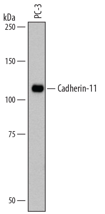 Detection of Human Cadherin‑11 by Western Blot.Western blot shows lysates of PC‑3 human prostate cancer cell line. PVDF membrane was probed with 1 µg/mL of Mouse Anti-Human Cadherin‑11 Monoclonal Antibody (Catalog # MAB1790) followed by HRP-conjugated Anti-Mouse IgG Secondary Antibody (Catalog # HAF007). A specific band was detected for Cadherin‑11 at approximately 110 kDa (as indicated). This experiment was conducted under reducing conditions and using Immunoblot Buffer Group 1.
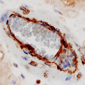 Cadherin‑11 in Human Placenta.Cadherin‑11 was detected in immersion fixed paraffin-embedded sections of human placenta using Mouse Anti-Human Cadherin‑11 Monoclonal Antibody (Catalog # MAB1790) at 8 µg/mL overnight at 4 °C. Tissue was stained using the Anti-Mouse HRP-DAB Cell & Tissue Staining Kit (brown; Catalog # CTS002) and counterstained with hematoxylin (blue). View our protocol for Chromogenic IHC Staining of Paraffin-embedded Tissue Sections.
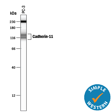 Detection of Human Cadherin‑11 by Simple WesternTM. Simple Western lane view shows lysates of PC‑3 human prostate cancer cell line, loaded at 0.2 mg/mL. Specific bands were detected for Cadherin‑11 at approximately 114 & 137 kDa (as indicated) using 20 µg/mL of Mouse Anti-Human Cadherin‑11 Monoclonal Antibody (Catalog # MAB1790). This experiment was conducted under reducing conditions and using the 12-230 kDa separation system. Non-specific interaction with the 230 kDa Simple Western standard may be seen with this antibody.
Product References
N-cadherin-mediated cell-cell adhesion promotes cell migration in a - N-cadherin-mediated cell-cell adhesion promotes cell migration in a
Shih W, Yamada S. J Cell Sci. 2012 Aug 1;125(Pt 15):3661-70. Epub 2012 Mar 30.
Hypoxia-induced abrogation of contact-dependent inhibition of rheumatoid - Hypoxia-induced abrogation of contact-dependent inhibition of rheumatoid
Nonomura Y, Mizoguchi F, Suzuki A, Nanki T, Kato H, Miyasaka N, Kohsaka H. J Rheumatol. 2009 Apr;36(4):698-705.