![All lanes : Anti-COX5A antibody [EPR14207(B)] (ab181226) at 1/10000 dilutionLane 1 : HepG2 cell lysateLane 2 : HeLa cell lysateLane 3 : A431 cell lysateLysates/proteins at 20 µg per lane.SecondaryGoat Anti-Rabbit IgG, (H+L), Peroxidase conjugate at 1/1000 dilution](http://www.bioprodhub.com/system/product_images/ab_products/2/sub_2/1454_ab181226-214619-ab181226WB.jpg)
All lanes : Anti-COX5A antibody [EPR14207(B)] (ab181226) at 1/10000 dilutionLane 1 : HepG2 cell lysateLane 2 : HeLa cell lysateLane 3 : A431 cell lysateLysates/proteins at 20 µg per lane.SecondaryGoat Anti-Rabbit IgG, (H+L), Peroxidase conjugate at 1/1000 dilution
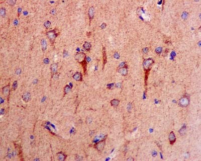
Immunohistochemical analysis of paraffin-embedded Human brain tissue labeling COX5A with ab181226 at 1/250 dilution followed by prediluted HRP Polymer for Rabbit IgG. Counter stained with Hematoxylin.
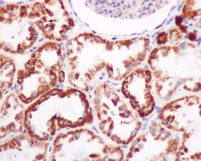
Immunohistochemical analysis of paraffin-embedded Human kidney tissue labeling COX5A with ab181226 at 1/100 dilution followed by prediluted HRP Polymer for Rabbit IgG. Counter stained with Hematoxylin.
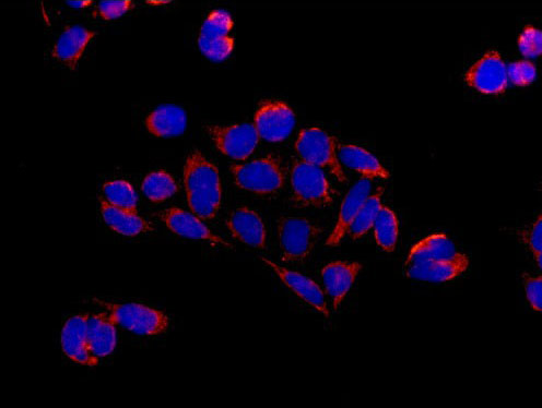
Immunofluorescent analysis of acetone-fixed HeLa cells labeling COX5A with ab181226 at 1/250 dilution, followed by Goat anti rabbit IgG (Alexa Fluor® 555) secondary antibody at 1/200 dilution (red). Counter stained with Dapi (blue).
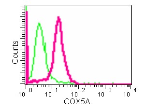
Flow cytometric analysis of 2% paraformaldehyde-fixed HeLa cells labeling COX5A with ab181226 at 1/10 dilution (red) compared to a Rabbit monoclonal IgG isotype control (green), followed by Goat anti rabbit IgG (FITC) secondary antibody at 1/150 dilution.
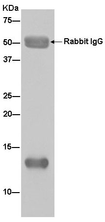
Western blot analysis of HeLa cell lysate immunoprecipitated with ab181226 at 1/50 dilution.Secondary: Goat Anti-Rabbit IgG, (H+L), Peroxidase conjugate at 1/1000 dilution.