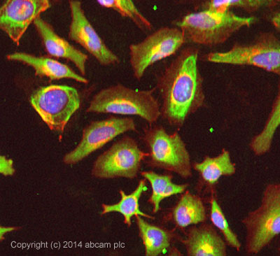
ICC/IF image of ab155204 stained HeLa cells. The cells were 100% methanol fixed (5 min) and then incubated in 1%BSA / 10% normal goat serum / 0.3M glycine in 0.1% PBS-Tween for 1h to permeabilise the cells and block non-specific protein-protein interactions. The cells were then incubated with the antibody ab155204 at 5µg/ml overnight at +4°C. The secondary antibody (pseudo-colored green) was Dylight® 488 goat anti- rabbit (ab96899) IgG (H+L) preadsorbed, used at a 1/250 dilution for 1h. Alexa Fluor® 594 WGA was used to label plasma membranes (pseudo-colored red) at a 1/200 dilution for 1h at room temperature. DAPI was used to stain the cell nuclei (pseudo-colored blue) at a concentration of 1.43µM for 1hour at room temperature.
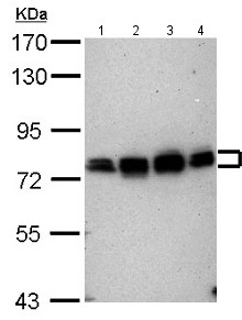
All lanes : Anti-CPEB4 antibody - N-terminal (ab155204) at 1/10000 dilutionLane 1 : NT2D1 whole cell lysateLane 2 : PC3 whole cell lysateLane 3 : U87-MG whole cell lysateLane 4 : Sk-N-SH whole cell lysateLysates/proteins at 30 µg per lane.
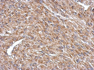
Immunohistochemical analysis of paraffin embedded U87 xenograft tissue labeling CPEB4 with ab155204 at 1/500 dilution.