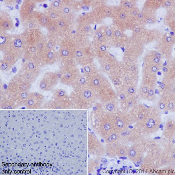
Immunohistochemical analysis of paraffin-embedded Human liver tissue labeling Cytochrome P450 2D6 with ab185625 at 1/500 dilution, followed by Goat Anti-Rabbit IgG H&L (HRP) (ab97051) secondary antibody at 1/500 dilution. Cytoplasmic staining on Human liver is observed. Counter stained with Hematoxylin.Negative control: Used PBS instead of primary antibody, secondary antibody is Goat Anti-Rabbit IgG H&L (HRP) (ab97051) at 1/500 dilution.
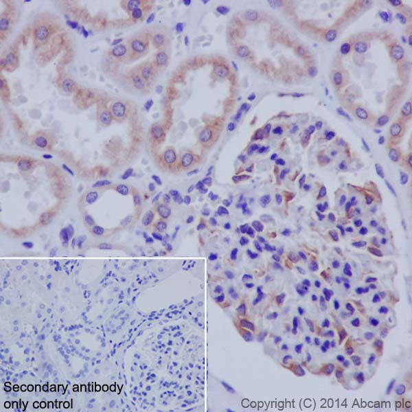
Immunohistochemical analysis of paraffin-embedded Human kidney tissue labeling Cytochrome P450 2D6 with ab185625 at 1/500 dilution, followed by Goat Anti-Rabbit IgG H&L (HRP) (ab97051) secondary antibody at 1/500 dilution. Cytoplasmic staining on Human kidney is observed. Counter stained with Hematoxylin.Negative control: Used PBS instead of primary antibody, secondary antibody is Goat Anti-Rabbit IgG H&L (HRP) (ab97051) at 1/500 dilution.
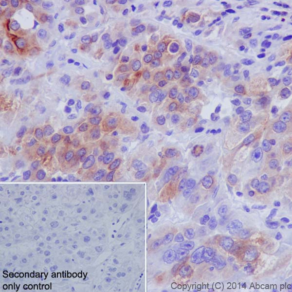
Immunohistochemical analysis of paraffin-embedded Human hepatocellular carcinoma tissue labeling Cytochrome P450 2D6 with ab185625 at 1/500 dilution, followed by Goat Anti-Rabbit IgG H&L (HRP) (ab97051) secondary antibody at 1/500 dilution. Cytoplasmic staining on Human hepatocellular carcinoma is observed. Counter stained with Hematoxylin.Negative control: Used PBS instead of primary antibody, secondary antibody is Goat Anti-Rabbit IgG H&L (HRP) (ab97051) at 1/500 dilution.
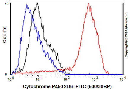
Flow cytometric analysis of 2% paraformaldehyde-fixed HT-29 (Human colorectal adenocarcinoma cells) cells labeling Cytochrome P450 2D6 with ab185625 at 1/300 dilution (red) compared with a rabbit monoclonal IgG isotype control (ab172730) (black) and an unlabelled control (cells without incubation with primary antibody and secondary antibody; blue). Goat anti rabbit IgG (FITC) at 1/150 dilution was used as the secondary antibody.
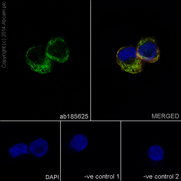
Immunofluorescent analysis of 4% paraformaldehyde-fixed, 0.1% Triton X-100 permeabilized HT-29 (Human colorectal adenocarcinoma cells) cells labeling Cytochrome P450 2D6 with ab185625 at 1/1000 dilution, followed by Goat anti-rabbit IgG (Alexa Fluor® 488) (ab150077) secondary antibody at 1/400 dilution (green). Cytoplasmic staining on HT-29 cell line is observed. The nuclear counter stain is DAPI (blue). Tubulin is detected with ab7291 (anti-Tubulin mouse mAb) at 1/500 dilution and ab150120 (AlexaFluor®594 Goat anti-Mouse secondary) at 1/500 dilution (red).The negative controls are as follows:-ve control 1: ab185625 at 1/1000 dilution followed by ab150120 (AlexaFluor®594 Goat anti-Mouse secondary) at 1/500 dilution.-ve control 2: ab7291 (anti-Tubulin mouse mAb) at 1/500 dilution followed by ab150077 (Alexa Fluor®488 Goat Anti-Rabbit IgG H&L) at 1/400 dilution.
![All lanes : Anti-Cytochrome P450 2D6 antibody [EPR17868] (ab185625) at 1/10000 dilutionLane 1 : Full-length Cytochrome P450 2D6 transfected 293T lysateLane 2 : Non-transfected 293T lysateLysates/proteins at 10 µg per lane.SecondaryAnti-Rabbit IgG (HRP), specific to the non-reduced form of IgG at 1/1000 dilution](http://www.bioprodhub.com/system/product_images/ab_products/2/sub_2/4908_ab185625-238897-185625WBa.jpg)
All lanes : Anti-Cytochrome P450 2D6 antibody [EPR17868] (ab185625) at 1/10000 dilutionLane 1 : Full-length Cytochrome P450 2D6 transfected 293T lysateLane 2 : Non-transfected 293T lysateLysates/proteins at 10 µg per lane.SecondaryAnti-Rabbit IgG (HRP), specific to the non-reduced form of IgG at 1/1000 dilution
![Anti-Cytochrome P450 2D6 antibody [EPR17868] (ab185625) at 1/10000 dilution + Human fetal liver lysate at 20 µgSecondaryAnti-Rabbit IgG (HRP), specific to the non-reduced form of IgG at 1/1000 dilution](http://www.bioprodhub.com/system/product_images/ab_products/2/sub_2/4909_ab185625-238896-185625WBb.jpg)
Anti-Cytochrome P450 2D6 antibody [EPR17868] (ab185625) at 1/10000 dilution + Human fetal liver lysate at 20 µgSecondaryAnti-Rabbit IgG (HRP), specific to the non-reduced form of IgG at 1/1000 dilution
![All lanes : Anti-Cytochrome P450 2D6 antibody [EPR17868] (ab185625) at 1/10000 dilutionLane 1 : HeLa (Human epithelial cells from cervix adenocarcinoma) whole cell lysateLane 2 : K562 (Human chronic myelogenous leukemia cells from bone marrow) whole cell lysateLane 3 : HepG2 (Human liver hepatocellular carcinoma) whole cell lysateLysates/proteins at 20 µg per lane.SecondaryAnti-Rabbit IgG (HRP), specific to the non-reduced form of IgG at 1/1000 dilution](http://www.bioprodhub.com/system/product_images/ab_products/2/sub_2/4910_ab185625-238895-185625WBc.jpg)
All lanes : Anti-Cytochrome P450 2D6 antibody [EPR17868] (ab185625) at 1/10000 dilutionLane 1 : HeLa (Human epithelial cells from cervix adenocarcinoma) whole cell lysateLane 2 : K562 (Human chronic myelogenous leukemia cells from bone marrow) whole cell lysateLane 3 : HepG2 (Human liver hepatocellular carcinoma) whole cell lysateLysates/proteins at 20 µg per lane.SecondaryAnti-Rabbit IgG (HRP), specific to the non-reduced form of IgG at 1/1000 dilution
![Anti-Cytochrome P450 2D6 antibody [EPR17868] (ab185625) at 1/1000 dilution + HT-29 (Human colorectal adenocarcinoma cells) whole cell lysate at 10 µgSecondaryAnti-Rabbit IgG (HRP), specific to the non-reduced form of IgG at 1/1000 dilution](http://www.bioprodhub.com/system/product_images/ab_products/2/sub_2/4911_ab185625-238894-185625WBd.jpg)
Anti-Cytochrome P450 2D6 antibody [EPR17868] (ab185625) at 1/1000 dilution + HT-29 (Human colorectal adenocarcinoma cells) whole cell lysate at 10 µgSecondaryAnti-Rabbit IgG (HRP), specific to the non-reduced form of IgG at 1/1000 dilution