![Anti-DGAT1 antibody [EPR13430-4] - N-terminal (ab181180) at 1/50000 dilution + HeLa cell lysate at 10 µgSecondaryGoat Anti-Rabbit IgG, (H+L), Peroxidase conjugated at 1/1000 dilution](http://www.bioprodhub.com/system/product_images/ab_products/2/sub_2/8026_ab181180-214681-ab181180WB1.jpg)
Anti-DGAT1 antibody [EPR13430-4] - N-terminal (ab181180) at 1/50000 dilution + HeLa cell lysate at 10 µgSecondaryGoat Anti-Rabbit IgG, (H+L), Peroxidase conjugated at 1/1000 dilution
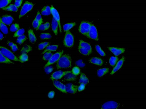
Immunofluorescence analysis of acetone-fixed HeLa cells, staining DGAT1 (green) with ab181180 at 1/100 dilution. Alexa Fluor®488-conjugated goat anti rabbit IgG was used as a secondary antibody at 1/200 dilution. Nuclei were counterstained with DAPI (blue).
![Anti-DGAT1 antibody [EPR13430-4] - N-terminal (ab181180) at 1/10000 dilution + Human fetal kidney lysate at 10 µgSecondaryGoat Anti-Rabbit IgG H&L (HRP) (ab136636) at 1/500 dilution](http://www.bioprodhub.com/system/product_images/ab_products/2/sub_2/8028_ab181180-214683-ab181180WB2.jpg)
Anti-DGAT1 antibody [EPR13430-4] - N-terminal (ab181180) at 1/10000 dilution + Human fetal kidney lysate at 10 µgSecondaryGoat Anti-Rabbit IgG H&L (HRP) (ab136636) at 1/500 dilution
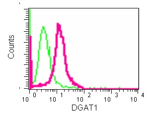
Flow cytometry analysis of DGAT1 expression in HeLa cells using ab181180 at 1/70 dilution (red) and a rabbit IgG as negative control (green).
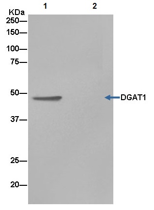
Western blot analysis on immunoprecipitation pellet from Human fetal kidney lysate (lane 1) or negative control (lane 2), labeling DGAT1 immunoprecipitated using ab181180 at 1/1500 dilution and HRP-conjugated anti-rabbit IgG preferentially detecting the non-reduced form of rabbit IgG.