")
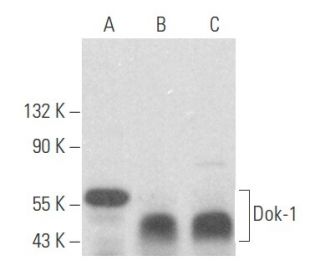
Dok-1 Antibody (A-3): sc-6929
- Dok-1 Antibody (A-3) is a mouse monoclonal IgG1 κ Dok-1 antibody, cited in 19 publications, provided at 200 µg/ml
- raised against amino acids 1-276 mapping at the N-terminus of Dok-1 of mouse origin
- Dok-1 Antibody (A-3) is recommended for detection of Dok-1 of mouse, rat and human origin by WB, IP, IF and IHC(P)
- Anti-Dok-1 Antibody (A-3) is available conjugated to agarose for IP; HRP for WB, IHC(P) and ELISA; and to either phycoerythrin or FITC for IF, IHC(P) and FCM
- also available conjugated to Alexa Fluor® 488, Alexa Fluor® 546, Alexa Fluor® 594 or Alexa Fluor® 647 for WB (RGB), IF, IHC(P) and FCM, and for use with RGB fluorescent imaging systems, such as iBright™ FL1000, FluorChem™, Typhoon, Azure and other comparable systems
- also available conjugated to Alexa Fluor® 680 or Alexa Fluor® 790 for WB (NIR), IF and FCM; for use with Near-Infrared (NIR) detection systems, such as LI-COR®Odyssey®, iBright™ FL1000, FluorChem™, Typhoon, Azure and other comparable systems
- Contact our Technical Service Department (or your local Distributor) for more information on how to receive a FREE 10 µg sample of Dok-1 (A-3): sc-6929.
- m-IgG Fc BP-HRP, m-IgG1 BP-HRP and m-IgGκ BP-HRP are the preferred secondary detection reagents for Dok-1 Antibody (A-3) for WB and IHC(P) applications. These reagents are now offered in bundles with Dok-1 Antibody (A-3) (see ordering information below).
QUICK LINKS
Dok-1 Antibody (A-3) is a mouse monoclonal IgG1 kappa light chain antibody that detects the Dok-1 protein of mouse, rat, and human origin by western blotting (WB), immunoprecipitation (IP), immunofluorescence (IF), and immunohistochemistry. Dok-1 (A-3) antibody is available in both non-conjugated and various conjugated forms, including agarose, horseradish peroxidase (HRP), phycoerythrin (PE), fluorescein isothiocyanate (FITC), and multiple Alexa Fluor® conjugates. Dok-1 plays a crucial role in cellular signaling pathways, particularly in the regulation of cell growth and differentiation, by acting as a docking protein that interacts with various signaling molecules. Dok-1′s ability to associate with the Ras GTPase-activating protein (Ras GAP) upon tyrosine phosphorylation is significant, as this association helps modulate the activity of Ras, a key player in cell proliferation and survival. Furthermore, Dok-1 is implicated in the signaling pathways activated by several growth factors, including platelet-derived growth factor (PDGF), vascular endothelial growth factor (VEGF), insulin, and insulin-like growth factor (IGF), highlighting Dok-1′s importance in both normal physiology and disease states, such as chronic myelogenous leukemia, where Dok-1 is a substrate of the constitutive tyrosine kinase activity of the p210 Bcr-Abl fusion protein. Additionally, Dok-1, along with other tyrosine kinase substrates like IRS-1 and Cas, contains multiple tyrosine residues and putative SH2 binding sites, which are essential for Dok-1′s function in signal transduction.
Alexa Fluor® is a trademark of Molecular Probes Inc., OR., USA
LI-COR® and Odyssey® are registered trademarks of LI-COR Biosciences
Dok-1 Antibody (A-3) References:
- Dok-1 and Dok-2 deficiency induces osteopenia via activation of osteoclasts. | Kawamata, A., et al. 2011. J Cell Physiol. 226: 3087-93. PMID: 21732353
- Dok-1 negatively regulates platelet integrin αIIbβ3 outside-in signalling and inhibits thrombosis in mice. | Niki, M., et al. 2016. Thromb Haemost. 115: 969-78. PMID: 26790499
- Dok-1 and Dok-2 Regulate the Formation of Memory CD8+ T Cells. | Laroche-Lefebvre, C., et al. 2016. J Immunol. 197: 3618-3627. PMID: 27664281
- Dok-1 regulates mast cell degranulation negatively through inhibiting calcium-dependent F-actin disassembly. | Du, H., et al. 2022. Clin Immunol. 238: 109008. PMID: 35421591
- Evidence that SH2 domains promote processive phosphorylation by protein-tyrosine kinases. | Mayer, BJ., et al. 1995. Curr Biol. 5: 296-305. PMID: 7780740
- Vascular endothelial cell growth factor promotes tyrosine phosphorylation of mediators of signal transduction that contain SH2 domains. Association with endothelial cell proliferation. | Guo, D., et al. 1995. J Biol Chem. 270: 6729-33. PMID: 7896817
- The IRS-1 signaling system. | Myers, MG., et al. 1994. Trends Biochem Sci. 19: 289-93. PMID: 8048169
- A 62-kilodalton tyrosine phosphoprotein constitutively present in primary chronic phase chronic myelogenous leukemia enriched lineage negative blast populations. | Wisniewski, D., et al. 1994. Leukemia. 8: 688-93. PMID: 8152267
- A Grb2-associated docking protein in EGF- and insulin-receptor signalling. | Holgado-Madruga, M., et al. 1996. Nature. 379: 560-4. PMID: 8596638
- p62(dok): a constitutively tyrosine-phosphorylated, GAP-associated protein in chronic myelogenous leukemia progenitor cells. | Carpino, N., et al. 1997. Cell. 88: 197-204. PMID: 9008160
- Identification of the Abl- and rasGAP-associated 62 kDa protein as a docking protein, Dok. | Yamanashi, Y. and Baltimore, D. 1997. Cell. 88: 205-11. PMID: 9008161
- Molecular cloning and characterization of p56dok-2 defines a new family of RasGAP-binding proteins. | Di Cristofano, A., et al. 1998. J Biol Chem. 273: 4827-30. PMID: 9478921
Ordering Information
| Product Name | Catalog # | UNIT | Price | Qty | FAVORITES | |
Dok-1 Antibody (A-3) | sc-6929 | 200 µg/ml | $316.00 | |||
Dok-1 Antibody (A-3): m-IgG Fc BP-HRP Bundle | sc-528168 | 200 µg Ab; 10 µg BP | $354.00 | |||
Dok-1 Antibody (A-3): m-IgGκ BP-HRP Bundle | sc-520478 | 200 µg Ab, 40 µg BP | $354.00 | |||
Dok-1 Antibody (A-3): m-IgG1 BP-HRP Bundle | sc-542796 | 200 µg Ab; 20 µg BP | $354.00 | |||
Dok-1 Antibody (A-3) AC | sc-6929 AC | 500 µg/ml, 25% agarose | $416.00 | |||
Dok-1 Antibody (A-3) HRP | sc-6929 HRP | 200 µg/ml | $316.00 | |||
Dok-1 Antibody (A-3) FITC | sc-6929 FITC | 200 µg/ml | $330.00 | |||
Dok-1 Antibody (A-3) PE | sc-6929 PE | 200 µg/ml | $343.00 | |||
Dok-1 Antibody (A-3) Alexa Fluor® 488 | sc-6929 AF488 | 200 µg/ml | $357.00 | |||
Dok-1 Antibody (A-3) Alexa Fluor® 546 | sc-6929 AF546 | 200 µg/ml | $357.00 | |||
Dok-1 Antibody (A-3) Alexa Fluor® 594 | sc-6929 AF594 | 200 µg/ml | $357.00 | |||
Dok-1 Antibody (A-3) Alexa Fluor® 647 | sc-6929 AF647 | 200 µg/ml | $357.00 | |||
Dok-1 Antibody (A-3) Alexa Fluor® 680 | sc-6929 AF680 | 200 µg/ml | $357.00 | |||
Dok-1 Antibody (A-3) Alexa Fluor® 790 | sc-6929 AF790 | 200 µg/ml | $357.00 |