")
FAS Antibody (B-10): sc-8009
- FAS Antibody (B-10) is a mouse monoclonal IgG1 κ FAS antibody, cited in 134 publications, provided at 200 µg/ml
- specific for an epitope mapping between amino acids 316-335 at the C-terminus of FAS of human origin
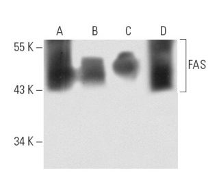
- FAS Antibody (B-10) is recommended for detection of FAS of human origin by WB, IP, IF, IHC(P), FCM and ELISA
- Anti-FAS Antibody (B-10) is available conjugated to agarose for IP; HRP for WB, IHC(P) and ELISA; and to either phycoerythrin or FITC for IF, IHC(P) and FCM
- also available conjugated to Alexa Fluor® 488, Alexa Fluor® 546, Alexa Fluor® 594 or Alexa Fluor® 647 for WB (RGB), IF, IHC(P) and FCM, and for use with RGB fluorescent imaging systems, such as iBright™ FL1000, FluorChem™, Typhoon, Azure and other comparable systems
- also available conjugated to Alexa Fluor® 680 or Alexa Fluor® 790 for WB (NIR), IF and FCM; for use with Near-Infrared (NIR) detection systems, such as LI-COR®Odyssey®, iBright™ FL1000, FluorChem™, Typhoon, Azure and other comparable systems
- Contact our Technical Service Department (or your local Distributor) for more information on how to receive a FREE 10 µg sample of FAS (B-10): sc-8009.
- m-IgG Fc BP-HRP, m-IgG1 BP-HRP and m-IgGκ BP-HRP are the preferred secondary detection reagents for FAS Antibody (B-10) for WB and IHC(P) applications. These reagents are now offered in bundles with FAS Antibody (B-10) (see ordering information below).
QUICK LINKS
SEE ALSO...
FAS Antibody (B-10) is a mouse monoclonal IgG1 kappa light chain antibody that specifically targets the FAS protein, also known as CD95, Fas cell surface death receptor, or TNFRSF6, in human samples. FAS mouse monoclonal antibody (B-10) can be utilized in various applications, including western blotting (WB), immunoprecipitation (IP), immunofluorescence (IF), immunohistochemistry (IHCP), flow cytometry (FCM), and enzyme-linked immunosorbent assay (ELISA). FAS protein plays a crucial role in regulating apoptosis, which is the process of programmed cell death that is essential for maintaining cellular homeostasis and immune system function. FAS protein mediates apoptosis to eliminate virus-infected or transformed cells, thereby contributing to the body′s defense mechanisms against malignancies and infections. FAS protein is a cell surface glycoprotein that belongs to a receptor family that includes CD40 and tumor necrosis factor receptors, and is expressed on a wide range of lymphoid cell lines. FAS protein interaction with FAS-L is vital for triggering the apoptotic signaling cascade, highlighting the importance of this pathway in immune responses. FAS monoclonal antibody (B-10) is available in both non-conjugated and various conjugated forms, including agarose, horseradish peroxidase, phycoerythrin, fluorescein isothiocyanate, and multiple Alexa Fluor® conjugates, making this reagent a valuable tool for researchers studying apoptosis and immune regulation.
Alexa Fluor® is a trademark of Molecular Probes Inc., OR., USA
LI-COR® and Odyssey® are registered trademarks of LI-COR Biosciences
FAS Antibody (B-10) References:
- Role of perforin in lymphocyte-mediated cytolysis. | Yagita, H., et al. 1992. Adv Immunol. 51: 215-42. PMID: 1502975
- Aging correlates with decreased beta-cell proliferative capacity and enhanced sensitivity to apoptosis: a potential role for Fas and pancreatic duodenal homeobox-1. | Maedler, K., et al. 2006. Diabetes. 55: 2455-62. PMID: 16936193
- Lack of UCP2 reduces Fas-mediated liver injury in ob/ob mice and reveals importance of cell-specific UCP2 expression. | Fülöp, P., et al. 2006. Hepatology. 44: 592-601. PMID: 16941708
- A central role of perforin in cytolysis? | Podack, ER., et al. 1991. Annu Rev Immunol. 9: 129-57. PMID: 1910674
- Perforin-dependent and -independent pathways of cytotoxicity mediated by lymphocytes. | Young, JD., et al. 1988. Immunol Rev. 103: 161-202. PMID: 3292393
- Mechanism of lymphocyte-mediated cytotoxicity. | Henkart, PA. 1985. Annu Rev Immunol. 3: 31-58. PMID: 3904772
- Molecular cloning and expression of the Fas ligand, a novel member of the tumor necrosis factor family. | Suda, T., et al. 1993. Cell. 75: 1169-78. PMID: 7505205
- Fas and its ligand in a general mechanism of T-cell-mediated cytotoxicity. | Hanabuchi, S., et al. 1994. Proc Natl Acad Sci U S A. 91: 4930-4. PMID: 7515183
- The Fas protein is expressed at high levels on CD4+CD8+ thymocytes and activated mature lymphocytes in normal mice but not in the lupus-prone strain, MRL lpr/lpr. | Drappa, J., et al. 1993. Proc Natl Acad Sci U S A. 90: 10340-4. PMID: 7694292
Ordering Information
| Product Name | Catalog # | UNIT | Price | Qty | FAVORITES | |
FAS Antibody (B-10) | sc-8009 | 200 µg/ml | $316.00 | |||
FAS Antibody (B-10): m-IgG Fc BP-HRP Bundle | sc-528198 | 200 µg Ab; 10 µg BP | $354.00 | |||
FAS Antibody (B-10): m-IgGκ BP-HRP Bundle | sc-520520 | 200 µg Ab, 40 µg BP | $354.00 | |||
FAS Antibody (B-10): m-IgG1 BP-HRP Bundle | sc-542811 | 200 µg Ab; 20 µg BP | $354.00 | |||
FAS Antibody (B-10) AC | sc-8009 AC | 500 µg/ml, 25% agarose | $416.00 | |||
FAS Antibody (B-10) HRP | sc-8009 HRP | 200 µg/ml | $316.00 | |||
FAS Antibody (B-10) FITC | sc-8009 FITC | 200 µg/ml | $330.00 | |||
FAS Antibody (B-10) PE | sc-8009 PE | 200 µg/ml | $343.00 | |||
FAS Antibody (B-10) Alexa Fluor® 488 | sc-8009 AF488 | 200 µg/ml | $357.00 | |||
FAS Antibody (B-10) Alexa Fluor® 546 | sc-8009 AF546 | 200 µg/ml | $357.00 | |||
FAS Antibody (B-10) Alexa Fluor® 594 | sc-8009 AF594 | 200 µg/ml | $357.00 | |||
FAS Antibody (B-10) Alexa Fluor® 647 | sc-8009 AF647 | 200 µg/ml | $357.00 | |||
FAS Antibody (B-10) Alexa Fluor® 680 | sc-8009 AF680 | 200 µg/ml | $357.00 | |||
FAS Antibody (B-10) Alexa Fluor® 790 | sc-8009 AF790 | 200 µg/ml | $357.00 | |||
FAS (B-10) Neutralizing Peptide | sc-8009 P | 100 µg/0.5 ml | $68.00 |