CHRNA1

Gene Symbol CHRNA1
Entrez Gene 395608
Alt Symbol NACHRA1
Species Chicken
Gene Type protein-coding
Description cholinergic receptor, nicotinic, alpha 1 (muscle)
Other Description acetylcholine receptor subunit alpha|alpha-1 subunit, nicotinic acetylcholine receptor|cholinergic receptor, nicotinic, alpha polypeptide 1 (muscle)
Swissprots P09479
Accessions AAA48564 AAA48565 AAC06012 CAA30282 P09479 AJ250359 CAB59624 BX932014 NM_204816 NP_990147
Function After binding acetylcholine, the AChR responds by an extensive change in conformation that affects all subunits and leads to opening of an ion-conducting channel across the plasma membrane.
Subcellular Location Cell junction, synapse, postsynaptic cell membrane; Multi-pass membrane protein. Cell membrane; Multi-pass membrane protein.
Top Pathways Neuroactive ligand-receptor interaction

Anti-Nicotinic Acetylcholine Receptor alpha 1 antibody - ab28489 from Abcam

Prices $370.00
Sizes 100 µl
Host Rabbit
Clonality Polyclonal
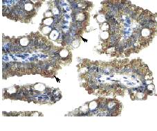
Immunohistochemical analysis of Nicotinic Acetylcholine Receptor alpha expression in paraffin embedded human intestine tissue section using 4 µg/ml ab28489. Arrows indicate positively labelled epithelial cells of intestinal villus.

CHRNA1 Monoclonal Antibody (88B) - MA3-043 from Thermo Fisher Scientific

Prices $375.00
Sizes 100 µl
Host Mouse
Clonality Monoclonal
Clone 88B
Immunohistochemistry was performed on normal biopsies of deparaffinized Mouse kidney tissue. To expose target proteins, heat induced antigen retrieval was performed using 10mM sodium citrate (pH6.0) buffer, microwaved for 8-15 minutes. Following antigen retrieval tissues were blocked in 3% BSA-PBS for 30 minutes at room temperature. Tissues were then probed at a dilution of 1:20 with a mouse monoclonal antibody recognizing Nicotinic Acetylcholine Receptor (MA3-043) or without primary antibody (negative control) overnight at 4°C in a humidified chamber. Tissues were washed extensively with PBST and endogenous peroxidase activity was quenched with a peroxidase suppressor. Detection was performed using a biotin-conjugated secondary antibody and SA-HRP, followed by colorimetric detection using DAB. Tissues were counterstained with hematoxylin and prepped for mounting.
Search more